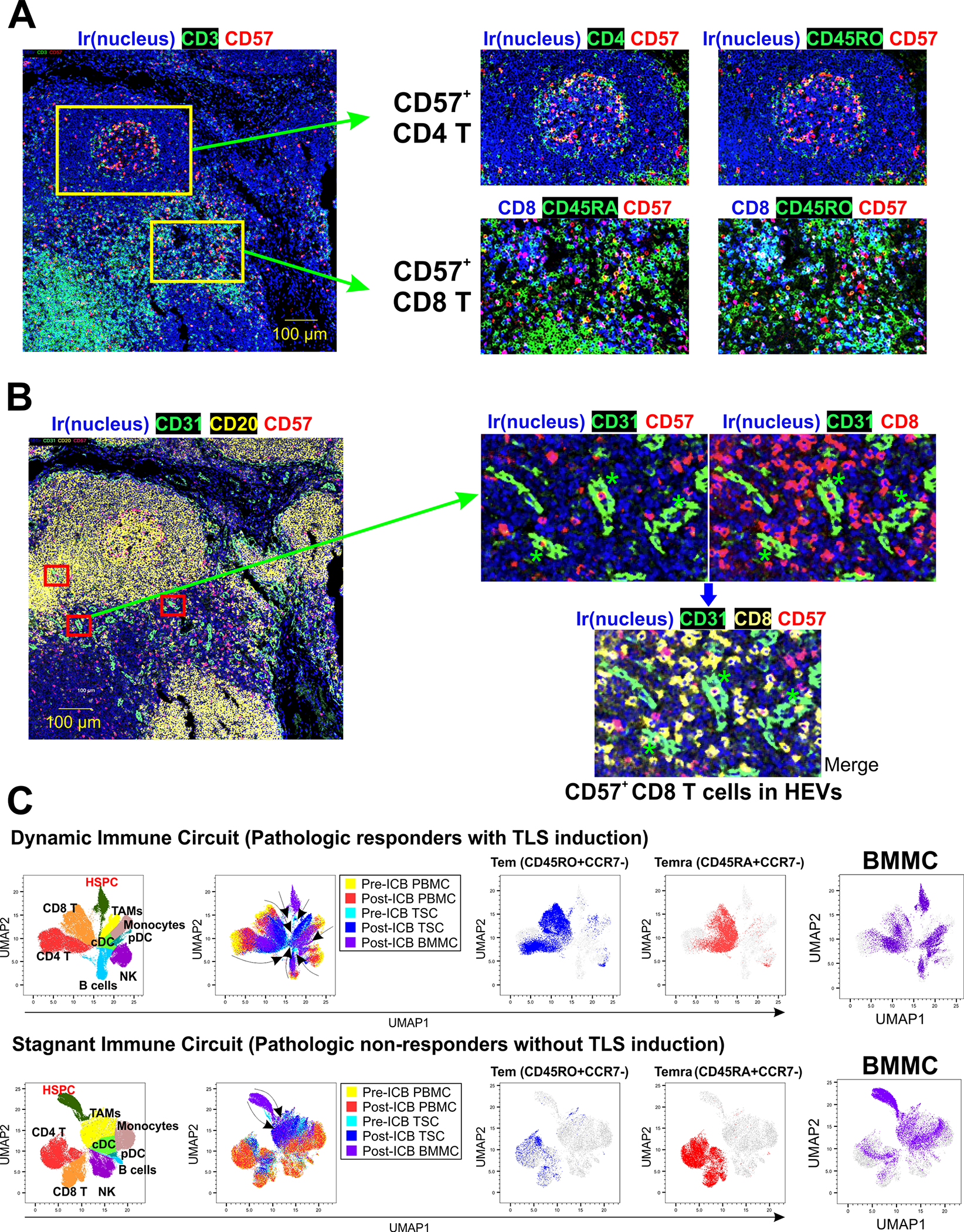
Figure 5. Interface of systemic and local anti-tumor immunity.

A. Identification of CD57+ T cells in the tumor immune environment after ICB. CD57+ T cells were located in germinal center areas and T cell zones of MPM TLSs. CD57+ T cells in germinal centers were CD45RO+ CD4 T cells. CD57+ T cells in T cell zones contained CD45RA+ and CD45RO+ CD8 T cells, consistent with CD8 Temra and CD8 Tem, respectively. B. Identification of CD57+ CD8 T cells in high endothelial venules (HEVs) expressing CD31 (marked with red rectangles in the lower magnification figure on the left, and with green asterisks on the higher magnification figure on the right). Higher magnification views demonstrate CD57+ T cells in HEVs. C. Cluster analysis of major cell types within the matched bone marrow, blood, and tumors was performed with CyTOF in 2 responders to ICB (PR and TLS formation) and 3 non-responders (no PR and no TLS formation). We observed a dynamic interaction between systemic and local immunity in responders (dynamic immune circuit), shown as the convergence of immune cell differentiation states from pre-ICB PBMC into post-ICB tumor cells, including CD8 and CD4 Tem and Temra; and HSPC differentiation into mature multilineage cells including Temra and Tem. In contrast, a “stagnant immune circuit” was observed in non-responders, with unchanged differentiation states of circulating and tumor-infiltrating immune cells after ICB, failure of Temra recruitment into the tumor, and abundant HSPC and myeloid cells in the bone marrow.
