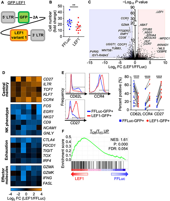
Figure 3: LEF1 overexpression promotes a central memory–like program in human NKTs.
A, Design of the GFP.LEF1 gammaretroviral construct for overexpression of LEF1 long isoform. B–F, NKTs underwent 10 days of primary expansion followed by restimulation with αGalCer-pulsed B-8-2 cells. They were then transduced with the GFP.LEF1 construct or GFP.FFLuc two days after secondary stimulation and phenotypes were studied eight days later. (B) Cell number was determined and fold change was calculated relative to input cell number. Mean ± SEM (n = 10 donors) are shown. **P < 0.01, paired Student’s t test. (C) GFP+ cells were FACS sorted from GFP.FFluc and GFP.LEF1 NKTs. RNA isolated from the sorted cells was processed for bulk RNAseq analysis. Volcano plot shows differential gene expression (LEF1/FFLuc) with P value cutoff at 0.05 and fold change ≤ −2 / ≥ 2. (D) Differentially expressed genes of interest from (C) were grouped by shared phenotype/function. Heat maps show fold change in expression (LEF1/FFLuc). Each column represents a donor with a total of six donors tested; false discovery rate (FDR) < 0.05 (FDR < 0.20 for PDCD1) in LEF1-GFP+ and FFLuc-GFP+ cells. (E) Surface expression of central memory markers on GFP+ cells from FFLuc- or LEF1-transduced NKTs were measured by flow cytometry. Representative donor histograms and paired percent positive (n ≥ 11 donors) are shown. **P < 0.01, ****P < 0.0001, paired Student’s t test for paired result from each donor. (F) GSEA plot showing enrichment for a central memory T–cell signature in LEF1-overexpressing NKTs.

