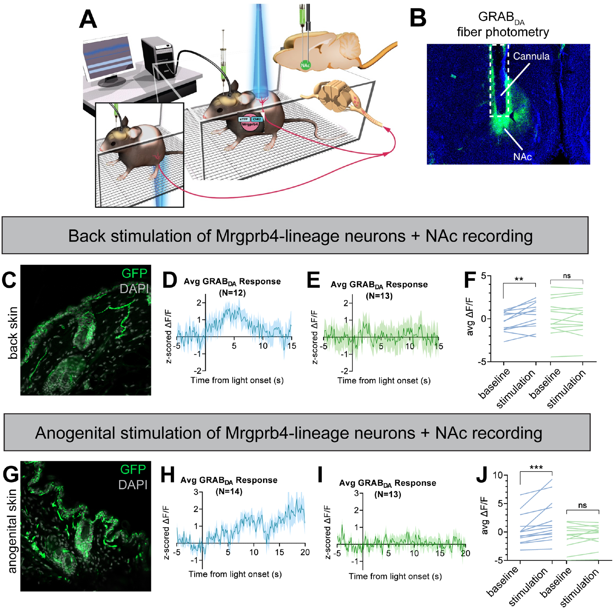
Figure 5: Activation of Mrgprb4-lineage neurons triggers dopamine release in the nucleus accumbens.

A) Schematic depicting the experimental setup. Female Mrgprb4Cre; RosaChR2/ChR2 mice with shaved backs, which had been injected with GRABDA to the NAc two weeks prior to testing, were placed under plastic chambers on a mesh platform. Pulsed blue (stimulating) or green (control) laser light (35mW, 10Hz sin wave) was shined to either the back skin (C-F) or skin surrounding the vagina (G-J) while recording GRABDA signals. B) Cannula placement and GRABDA expression in NAc C) ChR2-eYFP expression in Mrgprb4-lineage terminals in the back skin. D) Average GRABDA signal as z-score upon blue light (D) or green light (E) stimulation to the back. F) Average deltaF/F signals during pre-stimulation baseline (−5–0s) and during stimulation (0–6s) for each animal. Repeated measures two-way ANOVA revealed significant interaction between wavelength and time (p=0.0161) and Sidak’s multiple comparison test revealed significant difference in blue light (**p=0.0014) but not green light (p=0.9411) pre-stim compared to during stimulation. G-J) Average GRABDA signal as z-score upon blue light (H) or green light (I) stimulation to the anogenital region. J) Average deltaF/F signals during pre-stimulation baseline (−5–0s) and during stimulation (13–20s) for each animal. Repeated measures two-way ANOVA revealed significant interaction between wavelength and time (p=0.0110) and Sidak’s multiple comparison test revealed significant difference in blue light (***p=0.0003) but not green light (p=0.8547) pre-stim compared to during stimulation.
