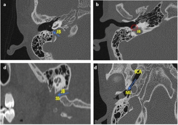
Figure 2.
Diagrammatic representation of all vascular variants on the axial and sagittal plane. JB jugular bulb, ICA internal carotid artery, ME middle-ear, SS sigmoid sinus. (a) HJB- blue arrow showing the bulb rising to the sigmoid plate; (b) JBD- red dotted circle showing absence of sigmoid plate due to descent of JB into the middle ear; (c) FJB- absence of the dome (rising bulb), blue arrow showing SS continue into the internal jugular vein; and (d) ICA-Deh: blue arrow showing focal dehiscence of the carotid canal into the middle ear.

