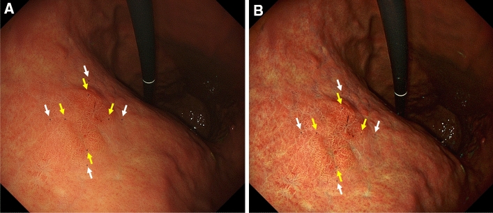
Figure 2.
Examples of actual endoscopic images used in the colorimetric evaluation process. A reddish depressed lesion is seen in the posterior wall of the middle gastric body. A total of 8 ROI were annotated on the surrounding non-neoplastic mucosa and EGC. ROIs were set at the same point on the WLI (A) and TXI (B). The yellow arrows indicate the EGC, and the white arrows indicate the surrounding non-neoplastic mucosa. All ROIs were standardized to 25 pixels (5 × 5).

