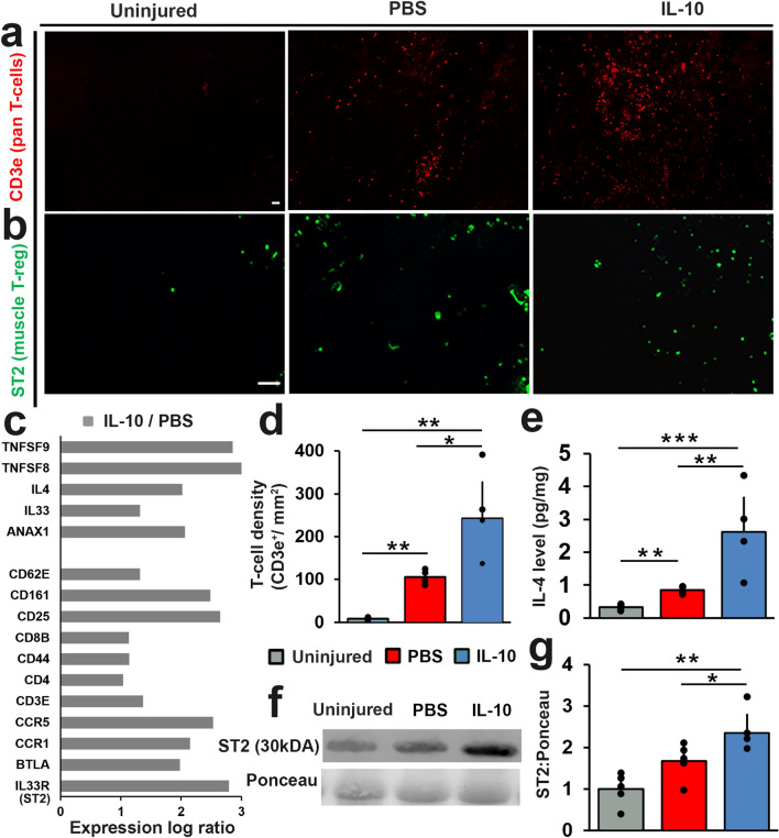
Figure 4.
IL-10 delivery boosted the infiltration of pro-regenerative T-regulatory cells at the site of VML repair. TA muscle cross-sections were stained for (a) CD3e (red) and (b) ST2 (green). Representative TA cross-sections from uninjured, PBS, and IL-10 are presented. Scale bar = 100 µm. (c) Key differentially expressed (FDR < 0.05) T-cell related genes are presented as log ratio (log2FC) by comparing the transcriptome of IL-10 treatment to PBS controls. (d) CD3e immunostained tissue cross-sections were used to quantify T-cells density (CD3e+/mm2). (e) IL-4 concentration (pg/mg) was determined from protein lysate using multiplexed ELISA. (f) Representative immunoblots stained with ST2 and Ponceau (original blot images shown in Fig. S3). (g) The relative (to Ponceau) ST2 protein contents for both IL-10 treatment and PBS controls were quantified from Western blots. ST2 protein concentration values were normalized to uninjured controls. Group means + SD are presented; n = 4–5/group. The *, **, and *** indicate statistically significant difference of p < 0.05, p < 0.01, and p < 0.001 between groups.

