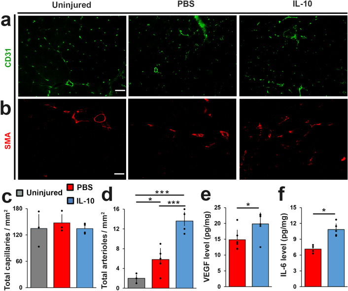
Figure 6.
IL-10 promoted repair site arteriole formation. TA muscle cross-sections prepared from 56 DPI were individually stained for (a) CD31 (green) or (b) α-SMA (red). Scale bar = 100 µm. Immuno-stained tissue cross sections prepared from 56 DPI tissue samples were quantified to determine (c) capillary density (capillaries/mm2), and (d) arteriole density (arterioles/mm2). (e) VEGF and (f) IL-6 concentrations (pg/mg) were measured from 14 DPI tissue samples using multiplex ELISA. Group means + SD are presented; n = 4–5/group. The *, **, and *** indicate statistically significant difference of p < 0.05, p < 0.01, and p < 0.001 between groups.

