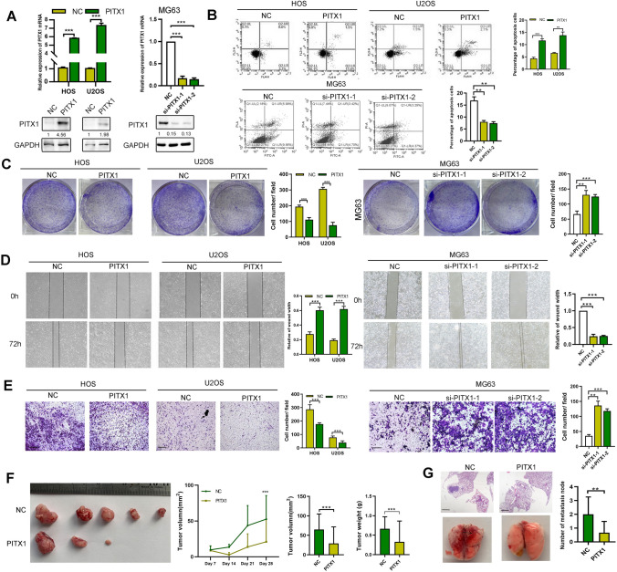
Fig. 1.
Function of PITX1 in OS in vitro and in vivo. A PITX1 expression was detected by qRT-PCR and western blotting following transient transfection of the PITX1 plasmid or two siRNAs in HOS, U2OS and MG63 cells. B Apoptosis of PITX1-overexpressing or -knockdown OS cells was measured by Annexin V-PI staining and flow cytometry. Percentage of apoptotic cells (Q2 + Q4) was determined after 48 h following PITX1 plasmid or siRNA transfection. C Colony formation of NC and PITX1-overexpressing or -knockdown OS cells was detected. D Wound healing assay was performed to measure the migration of NC and PITX1-overexpressing or -knockdown OS cells. E Transwell assays were performed to detect the migration of NC and PITX1-overexpressing or -knockdown OS cells. Scale bar, 100 μm. F Images of tumors obtained from NC or stable PITX1-overexpressing tumors of BALB/c nude mice after 28 days (n = 5 each group). Tumor volumes were measured every 7 days and the tumor weights were determined at the 28th day after inoculation. G The H&E staining images obtained from lung metastatic nodules from NC or PITX1-transfected tumor cells in BALB/c nude mice after 35 days (n = 5 each group). Scale bar, 400 μm. Data represent the mean ± SD of 3 separate determinations. *p < 0.05, **p < 0.01, ***p < 0.001 by Student’s t test

