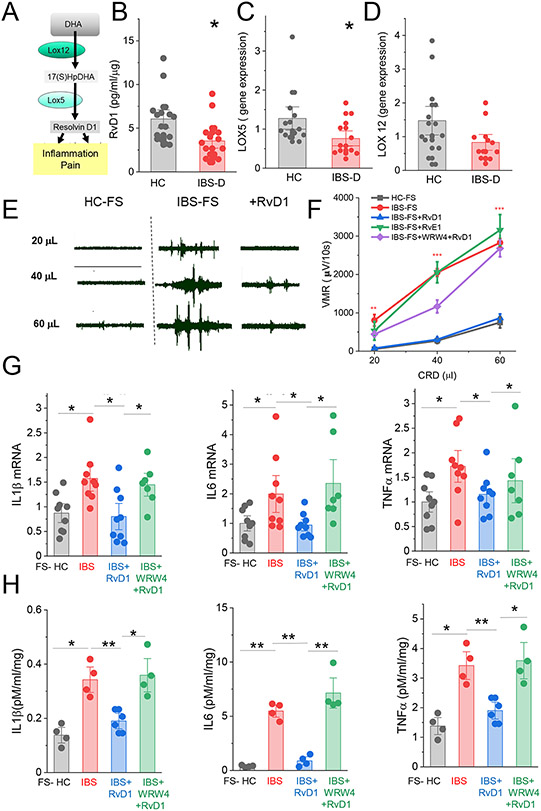
Fig 1.
Role of RvD1 in colon. (A) Schematic pathway of RvD1 synthesis. (B) ELISA data show the level of RvD1 in colonic biopsy samples from healthy control individuals (HC, n=16) and IBS-D patients (n=16). *, P<0.01. qPCR data showing relative expression of LOX5 mRNA (C), LOX12 mRNA (D) in colonic biopsy samples from HC or IBS-D patients. Data are expressed as means with SEM. *, P<0.05 from HC; unpaired t-test. (E) Representative recordings of VMR to colorectal distention after administration of fecal supernatant from HC (HC-FS, left panel) or fecal supernatant from IBS-D patient (IBS-FS, middle panel), and 30 minutes after administration of RvD1 (100ng/mouse ip, right panel). (F) Summary graph of VMR to colorectal distention shows that intracolonic administration of IBS-FS (n = 13) cause visceral hypersensitivity when compared to VMR generated in response to intracolonic administration of HC-FS (n=8). The enhanced VMR was abolished by i.p. administration of RvD1 (n = 7) but not RvE1 (1000 ng/mouse, n=6). The effect of RvD1 was inhibited by FPR2 receptor blocking peptide WRW4 (n = 6). (G) qPCR data show relative expression of mRNA for IL1b, IL-6, TNFα in colonic tissue from mice pretreated with HC-FS, IBS-FS, IBS-FS+RvD1 and IBS-FS+RvD1+WRW4. Each dot represents an individual mouse. Results are expressed as mean ± SEM; *, P<0.05 ANOVA with Bonferoni post-hock test. (H) ELISA data show relative expression of IL1b, IL-6, TNFα in supernatants from colonic tissue pretreated with HC-FS, IBS-FS, IBS-FS+RvD1 and IBS-FS+RvD1+WRW4. Each dot represents an individual mouse. Results are expressed as mean ± SEM; *, P<0.05 ANOVA with Bonferoni post-hock test.

