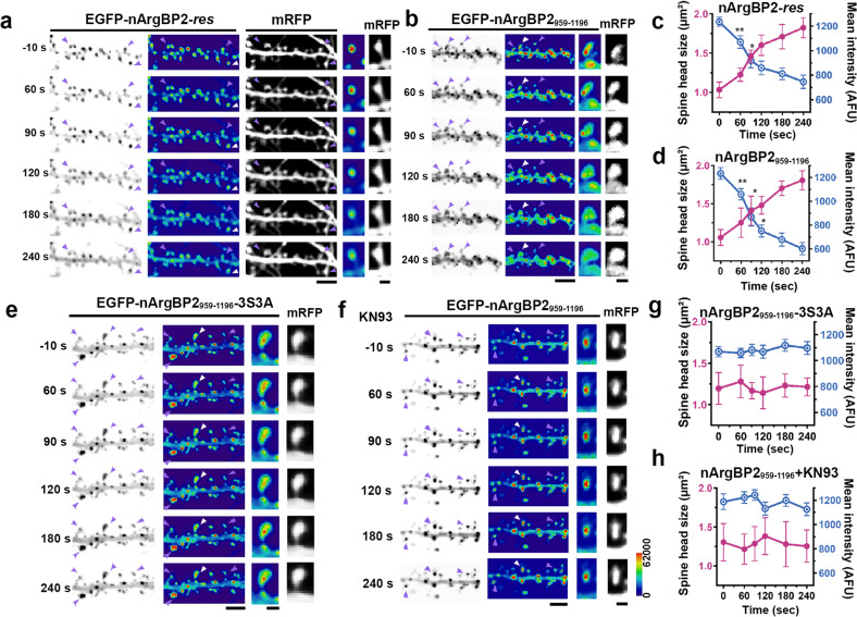
Fig. 2. nArgBP2 forms condensates in dendritic spines, and these condensates are dispersed by CaMKIIα-mediated phosphorylation during chemical-LTP, which spatiotemporally coincides with spine head enlargement.
Hippocampal neurons were transfected with EGFP-nArgBP2-res, EGFP-nArgBP2959-1196, or EGFP-nArgBP2959-1196-3S3A and mRFP-tagged shRNA-nArgBP2 to exclude the effect of endogenous nArgBP2. Representative time-lapse inverted images and pseudocolored images of hippocampal neurons transfected with EGFP-nArgBP2-res (a), EGFP-nArgBP2959-1196 (b), EGFP-nArgBP2959-1196-3S3A (e), or EGFP-nArgBP2959-1196 with KN93 treatment (f) upon cLTP stimulation. The third columns show magnified pseudocolored views of the spines indicated by white arrowheads. The last columns show the mRFP signal in the spine cytosol used as a volume marker. Scale bars: 10 and 1 μm, respectively. Plots of time-dependent changes in single spine head size and mean intensity of EGFP-nArgBP2-res (c), EGFP-nArgBP2959-1196 (d), EGFP-nArgBP2959-1196-3S3A (g), and EGFP-nArgBP2959-1196 with KN93 treatment (h) spines after cLTP. Both EGFP-nArgBP2-res and EGP-nArgBP2959-1196 but not EGFP-nArgBP2959-1196-3S3A and EGFP-nArgBP2959-1196 with KN93 treatment droplets in the dendritic spine dispersed rapidly during cLTP, leading to spine enlargement. See Methods for more details. n = 10; *p < 0.05; **p < 0.01; the spines and droplets in the third columns of a, b, e, and f were used for the analysis.

