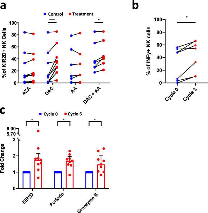
Fig. 6. Hypomethylating agents normalize the NK cell phenotype of MDS patients.
a Evaluation of KIR2D surface expression on NK cells of TET2/IDHMUT patients after treatment with azacitidine (AZA, n = 12), decitabine (DAC, n = 12), acid ascorbic (AA, n = 7), and DAC+AA (n = 7). Nonparametric two-sided Wilcoxon matched-pairs signed rank test was used to determine statistical significance, DAC: ***p = 0.001, DAC+AA: *p = 0.0156. b NK cells were isolated from patients’ PBMC before and after 3 cycles of treatment with AZA, and cultured overnight at 100U/ml of IL-2. Subsequently, cells were cultured with PMA-Ionomycin for 6 h. The frequency of responding cells in terms of IFN-γ was assessed by flow cytometry (n = 7). c KIR2D, perforin, and granzyme B expression were measured by flow cytometry in the blood of MDS patients (n = 9) before (in blue) and after (in red) 6 cycles of AZA treatment. Nonparametric two-sided Wilcoxon matched-pairs signed rank test was used to determine statistical significance, KIR2D: *p = 0.0195, Perforin: *p = 0.0273, Granzyme B: *p = 0.0273. Data are presented as medians and interquartile ranges. Source data are provided as a Source Data file.

