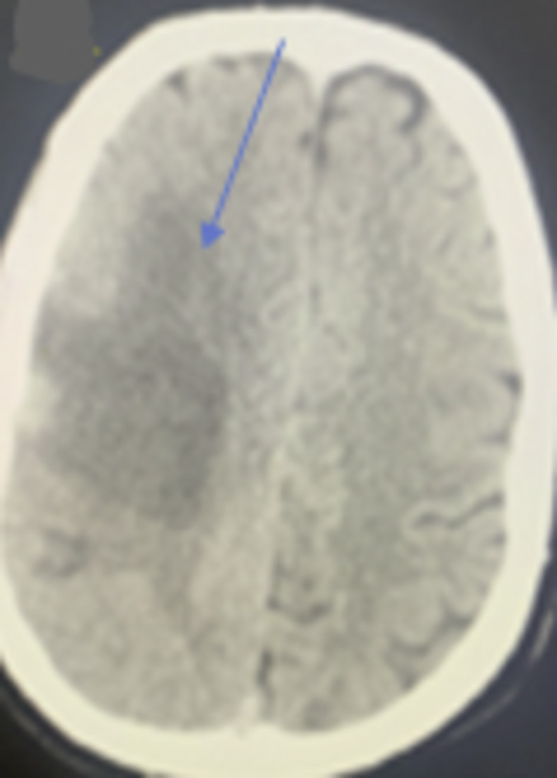
Fig. 4.

CT brain showed an ill-defined hypodense area in the right temporoparietal lobe with cortical and subcortical distribution in the region of the right middle cerebral artery and signs of hyperdense vessels associated with effacement of related cortical sulci (blue arrow).
