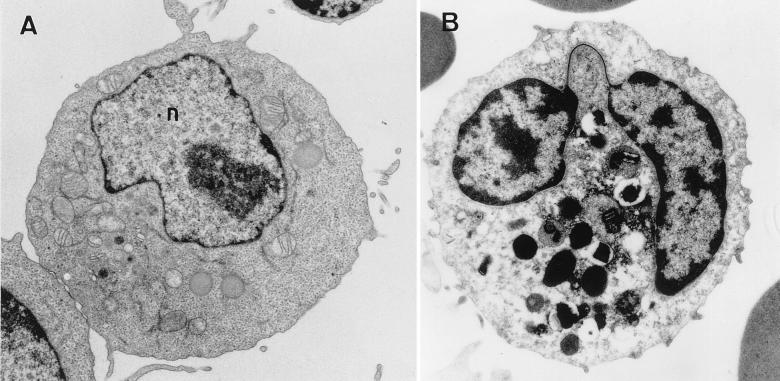
FIG. 3.
Electron micrograph of a stimulated lymphocyte (A) and a human granulocyte (B). (A) The nucleus (n) has many decondensed chromatin fibers throughout its volume. Condensed fibers are found at the nuclear periphery. (B) The highly lobed nucleus has a thin filament of chromatin connecting the lobes (arrow). Large amounts of condensed chromatin are found close to the nuclear envelope and in clumps internal to the nucleus. Magnification, ×5,000.

