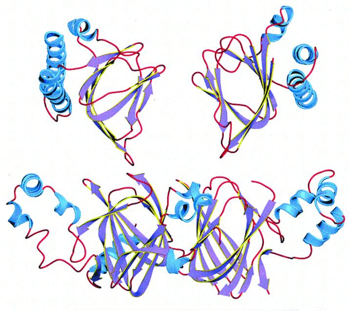
FIG. 2.
Comparative structures of two orientations of the arabinose-binding domain of the AraC protein (above) and the two-domain phaseolin storage protein (below), showing the similar β-barrel element in the center of each domain, with associated α-helixes. The apparent gap in the E/F loop in phaseolin is due to the lack of resolution of the 3D structure at that point (177).

