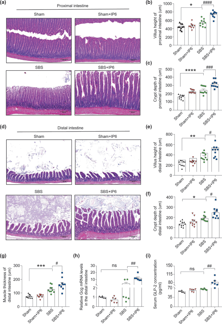
Fig. 2.
IP6 improves histological adaptation of the residual intestine in the SBS rats. (a) Representative images of hematoxylin-eosin (HE) staining of the proximal small intestine of rats in different groups (n = 10 per group). Scale bars, 250 μm. (b) Comparison of villus height in the proximal small intestine of rats in different groups (n = 10 per group). (c) Comparison of crypt depth in the proximal small intestine of rats in different groups (n = 10 per group). (d) Representative images of HE staining of the distal small intestine of rats in different groups (n = 10 per group). Scale bars, 250 μm. (e) Comparison of villus height in the distal small intestine of rats in different groups (n = 10 per group). (f) Comparison of crypt depth in the distal small intestine of rats in different groups (n = 10 per group). (g) Comparison of muscle thickness in the distal small intestine of rats in different groups (n = 10 per group). (h) The relative mRNA expression levels of Gcg of the mucosa in the distal small intestine (n = 6 per group). Values were normalized to Actb expression. (i) The levels of glucagon-like peptide-2 (GLP-2) in the serum were evaluated by ELISA analysis (n = 6 per group). Values are mean ± SEM; ns, not significant; * P < 0.05, ** P < 0.01, *** P < 0.001, **** P < 0.0001, SBS group versus Sham group; # P < 0.05, ## P < 0.01, ### P < 0.001, #### P < 0.0001, SBS + IP6 group versus SBS group.

