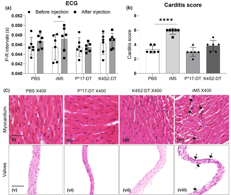
Fig. 2. Functional and pathological assessment of the heart.
Functional impairment of the heart before (•) and after injection of antigen (▪) was assessed by ECG (a). Statistical analysis performed by two-way ANOVA. Significance to before injection shown, *P < 0.05, **P < 0.01, ***P < 0.001. Development of carditis (b). Inflammatory changes in the myocardium and valvular tissue, characterised by mononuclear cell infiltration, were scored in PBS (•), rM5 (▪), P*17-DT (▴) and K4S2-DT (♦) treated rats. Statistical analysis performed by one-way ANOVA. Significance to PBS group shown, *P < 0.05, **P < 0.01, ***P < 0.001, ****P < 0.0001. Error bars represent standard deviation. Histological changes in cardiac tissues following exposure to PBS, P*17-DT, K4S2-DT and rM5 (c). Arrows indicates the possible mononuclear cell infiltration. Representative images from each group shown, scale bar represents 50 µm.

