Figure 5.

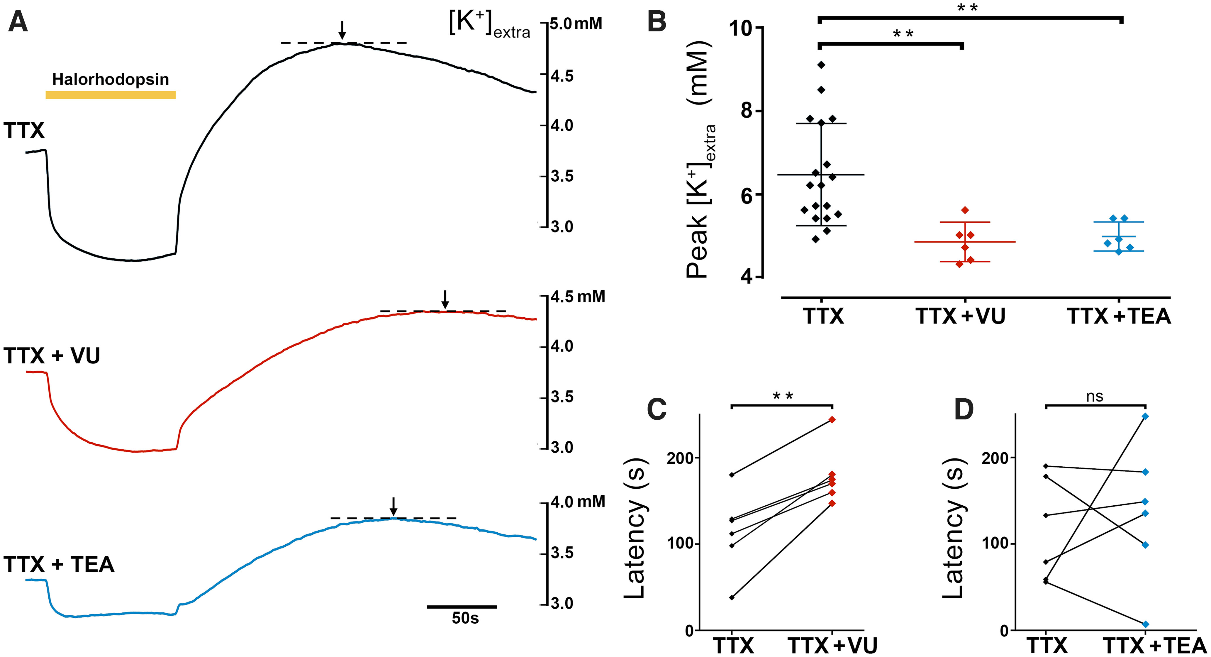
Halorhodopsin-induced K+ redistribution involves KCC2 and voltage-activated K+ channels. A, Recordings of [K+]extra during and after a 90 s period of halorhodopsin activation (yellow bar) in the presence of the Na+ channel blocker, TTX (blue, top), or the KCC2 blocker, VU0463271 (red, middle), or the blocker of voltage-gated K+ channels, TEA (black, bottom). B, The peak [K+]extra, after the end of illumination, for the three different drugs (p = 0.0037 for TTX vs TTX + VU and p = 0.0064 for TTX vs TTX + TEA, Kruskal–Wallis test with a Dunn's multiple comparisons test). Lines represent mean +/– std. C, The time to the peak [K+]extra, following a 90 s period of halorhodopsin activation, with and without the presence of the KCC2 blocker, VU0463271. Blockade of KCC2 causes a significant delay in the time to the peak (p = 0.0014, two-tailed paired t test). All experiments were performed in the presence of the Na+ channel blocker, TTX, to exclude any contribution to the [K+] fluxes made by rebound neuronal spiking. D, Similar measurements made with and without the voltage-gated K+ channel blocker, TEA, showing no significant effect.
