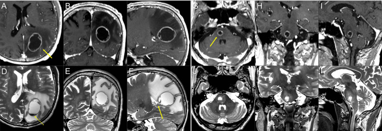
Figure 1. Magnetic resonance images for 10-fraction stereotactic radiosurgery planning.
The images show contrast-enhanced (CE) T1-weighted images (T1-WI) (A-C, G-I); T2-weighted images (T2-WI) (D-F, J-L); axial images (A, D, G, J); coronal images (B, E, H, K); and sagittal images (C, F, I, L).
(A-F) A cystic lesion (arrow in A) in the left parietal lobe is associated with massive surrounding edema and fluid sedimentation within the cyst (dashed arrow in D). The ventral side of the lesion is near the lateral ventricular wall (arrow in F). (G-I, J-L) A cystic lesion (arrow in G) in the pons with mild perilesional edema.

