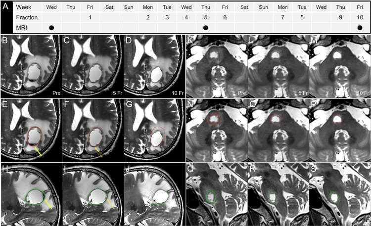
Figure 5. T2-weighted images obtained two days before stereotactic radiosurgery, at 5 and 10 fractions.
The images show (A) the actual schedule for fractionated SRS (fSRS) and MRI acquisition; (B-J) the parietal lesion; (K-S) the pons lesion; (B-G, K-P) axial images; (H-J, Q-S) sagittal images; (B, E, H, K, N, Q) before fSRS (pre); (C, F, I, L, O, R) at 5 fraction (fr); and (D, G, J, M, P, S) at 10 fr.
These T2-weighted image datasets were co-registered to the dedicated workstation, MIM MaestroTM (Cleveland, OH: MIM Software) and then fused to coincide with each other through the cranium. The pre-fSRS GTVs were contoured in red and green for the axial and sagittal images, respectively. In the parietal lesion, slight dorsomedial displacement of the lesion along with disappearance of the preexisting fluid sedimentation (arrows in E, H) was observed at 5 fr (dashed arrows in F, I). At 10 fr, a 39% (7.71 cm3) reduction in the GTV (19.93 to 12.22 cm3) and dorsomedial shifting of the tumor center were noted along with an improvement in the mass effect. Slight evisceration of the GTV dorsomedially from the initial dimensions was found ad eundem at 5 and 10 fr. In the pons lesion, no significant change was observed, apart from regression of the mild surrounding edema.

