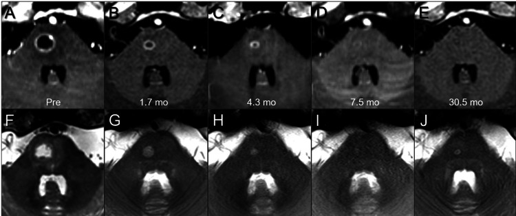
Figure 8. Serial magnetic resonance images for brain metastasis in the pons.
The images show (A-E) axial CE-T1-WI; axial T2-WI (F-J); before fSRS (pre) (A, F); at 1.7 months (mo) after fSRS (B, G); at 4.3 months (C, H); at 7.5 months (D, I); and at 30.5 months (E, J). These serial image datasets were co-registered and fused on MIM MaestroTM (Cleveland, OH: MIM Software).
CE: contrast-enhanced; T1-WI: T1-weighted image; T2-WI: T2-weighted image

