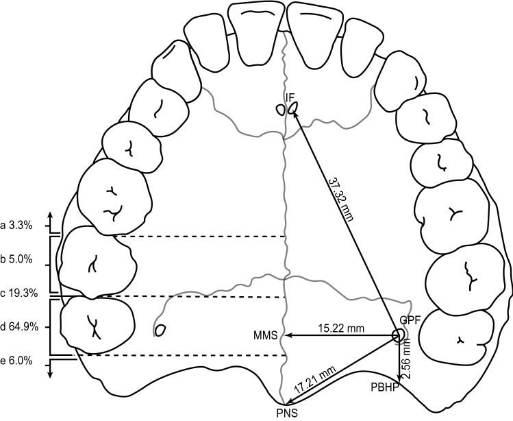
Fig. 3.
Illustration of the hard palate, displaying the greater palatine foramen in relation to anatomical landmarks and the maxillary molar teeth. The pooled mean distances from the greater palatine foramen (GPF) to four major anatomical landmarks (IF, MMS, PBHP, PNS) are shown on the right side of the diagram, while the pooled prevalence of the greater palatine foramen location in relation to the maxillary molar teeth are shown on the left side, (a–e). GPF greater palatine foramen, IF incisive foramen, MMS midline maxillary suture, PBHP posterior border of hard palate, PNS posterior nasal spine; a anterior to the mesial surface of the second maxillary molar; b opposite to the second maxillary molar; c between the second and the third maxillary molar; d opposite to the third maxillary molar; e distal to the third maxillary molar

