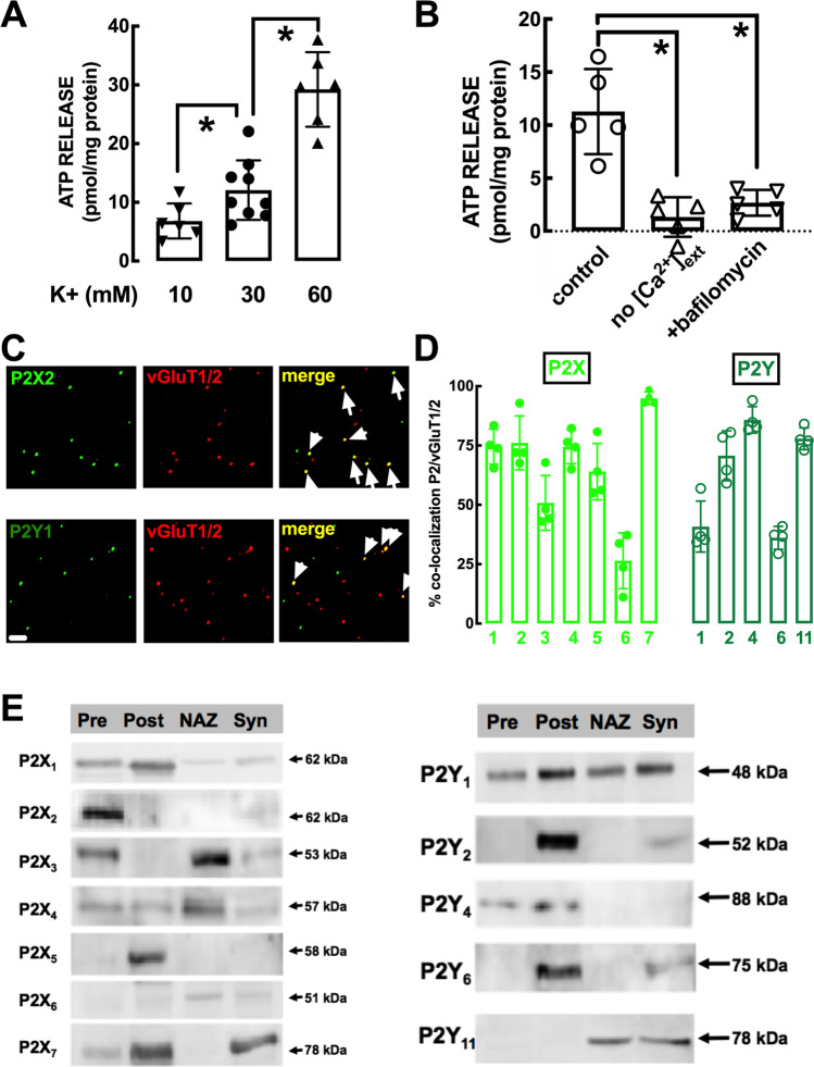
Fig. 2.
Intensity-dependent release of vesicular ATP and localization of different P2X and P2Y receptors in glutamatergic nerve terminals and within synapses in the mouse striatum. A The evoked release of ATP, evaluated with a luciferin-luciferase enzymatic assay, was triggered by exposure of mouse striatal synaptosomes to different concentrations of K+ (isomolar substitution of Na+ by K+ in the medium) and was larger at more intense stimulation. B ATP release was abolished in the absence of added extracellular calcium and inhibited in the presence of the vesicular proton pump inhibitor bafilomycin (100 nM), indicating a vesicular evoked release of ATP from nerve terminals. Data in A and B are mean ± S.E.M of 6–9 experiments (number of different animals tested); *p < 0.05 using a one-way ANOVA followed by Bonferroni’s post hoc test. C Representative photographs of immunocytochemistry staining of striatal nerve terminals with P2X2 receptor (upper row) and P2Y1 receptor subunit (lower row) and their co-localization with markers of glutamatergic nerve terminals (vesicular glutamate transporter type 1 and 2 – vGluT1/2) as highlighted by the arrows in the merged image. The scale bar is 10 μm. D Histograms representing the average co-localization of the different P2X receptor (P2XR) subunits or different P2YR in glutamatergic nerve terminals (i.e., labeled with vGluT1/2) from the mouse striatum. Data are mean ± S.E.M of 3 mice. E Subsynaptic localization in striatal synapses of the different P2XR subunits or different P2YR, assessed by Western blot analysis in the initial striatal synaptosomal preparation (Syn) and in purified extracts of the presynaptic active zone (Pre), the postsynaptic density (Post), and the extra-synaptic regions or non-active zone (NAZ). The blots are representative from two similar subsynaptic fractionations from the mouse striatum

