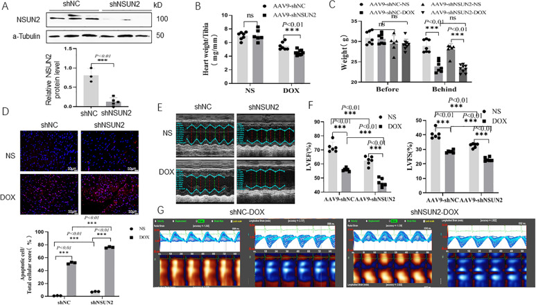
Fig. 2. Interfering with NSUN2 expression promoted DOX-induced myocardial injury.
A Western blot was used to detect the expression of NSUN2 protein in the heart tissue of mice treated with DOX (n = 3). B, C The weight and heart weight of mice in each group (n = 7). D TUNEL detected myocardial cell apoptosis in four groups of mice (n = 3, Bar = 50 µm). E M-mode ultrasonic representation of short-axis section. F Statistical chart of LVEF% and LVFS% of four groups of mice detected by small-animal ultrasound (n = 6). G Two-dimensional speckle tracking technology displays the diastolic phase (red) and systolic phase (blue) of the wall motion monitored by each tracer point in the order shown in the figure. The measurement data were shown as mean ± standard deviation and compared by Student’s t-test. *P < 0.05; **P < 0.01; ***P < 0.001.

