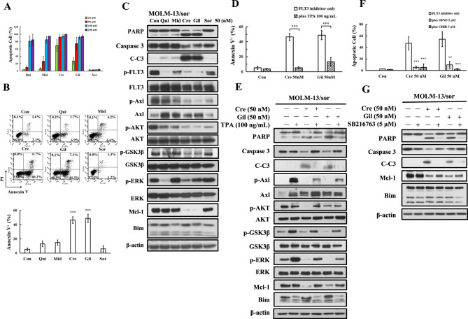
Fig. 4. Apoptosis induction and protein regulation of FLT3-ITD inhibitors in MOLM-13/sor cells.
A Apoptosis detected by AO-EB staining in MOLM-13/sor cells treated with each inhibitor for 24 h. B Apoptosis detected by Annexin V/PI staining in MOLM-13/sor cells treated by 50 nM of each inhibitor. C Protein regulation in MOLM-13/sor cells treated with each inhibitor at 50 nM for 24 h. D MOLM-13/sor cells pretreated with 100 ng/mL TPA and then with crenolanib and gilteritinib at 50 nM for 24 h. Apoptosis was detected by Annexin V/PI staining. E Apoptosis-related proteins analyzed by Western blot analysis. F MOLM-13/sor cells were pretreated with 5 μM GSK3β inhibitor CHIR-99021 or SB216763 for 4 h and then cotreated with crenolanib and gilteritinib for 24 h. Apoptotic cells were detected by AO-EB staining. G Protein regulation in MOLM-13/sor cells treated with SB216763 in combination with crenolanib and gilteritinib for 24 h. *P < 0.05, **P < 0.01, ***P < 0.001 compared with the FLT3 inhibitor group.

