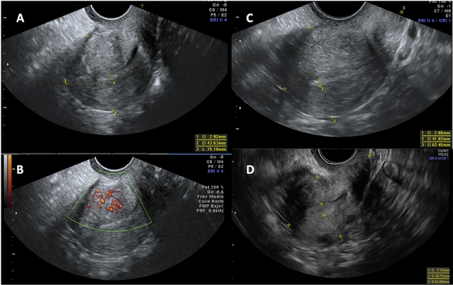
Figure 2.
Adenomyosis sonographic evolution at baseline and at 12 and 24 months of follow-up. (A and B) Baseline ultrasound with the presence of 5 adenomyosis criteria; (A) Hyperechoic islands, fan-shaped shadowing, uterine wall asymmetrical thickening, interrupted junctional zone and (B) Translesional vascularity; (C) 12-month follow-up with hyperechoic islands and uterine wall asymmetrical thickening as mild signs of adenomyosis. (D) 24-month follow-up with no signs of adenomyosis.

