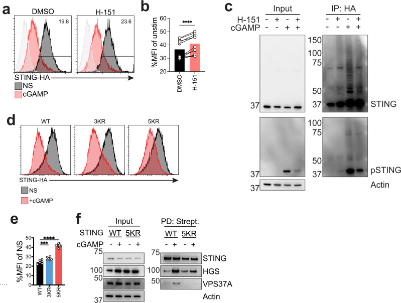
Fig. 7. STING oligomerization drives its ubiquitination and multiple lysines regulate STING degradation.
a mNG levels 293T STING-mNG stimulated with 4 µg/ml 2’3’-cGAMP(pS)2 (in medium) for 8 h with 1 µM H-151. One representative plot of n = 4 independent experiments with n = 3 technical replicates per experiment. b Ratio MFI of experiment same as in a. n = 4 independent experiments with n = 3 technical replicates per experiment. Each dot represents a replicate. Two-tailed paired t-test. c Immunoblot of the indicated proteins in THP-1 stably expressing HA-ubiquitin stimulated with 10 µg/ml cGAMP (in medium with digitonin) for 4 h. One experiment representative of n = 3 independent experiments. d HA intracellular staining in 293T stably expressing the indicated constructs stimulated with 2 µg/ml cGAMP (in perm buffer) for 6 h. One representative plot of n = 3 independent experiments with n = 2 technical replicates per experiment. e STING-HA MFI of cells as in d shown as %MFI of cGAMP stimulated over non-stimulated (NS) for each mutant. n = 3 independent experiments with n = 2 technical replicates per experiment. Each dot represents an individual replicate. One-way ANOVA with Dunnet multiple comparisons. f Immunoblot of the indicated proteins in input and streptavidin pulldown from 293T cells expressing STING WT or STING 5KR fused to TurboID stimulated with 2 µg/ml cGAMP (in perm buffer) for 3 h. One representative experiment of n = 3 independent experiments. In all panels, bar plots show mean and error bars standard deviation. Marker unit for Western blots is kDa. *p < 0.05, **p < 0.01, ***p < 0.001, ****p < 0.0001, ns not significant.

