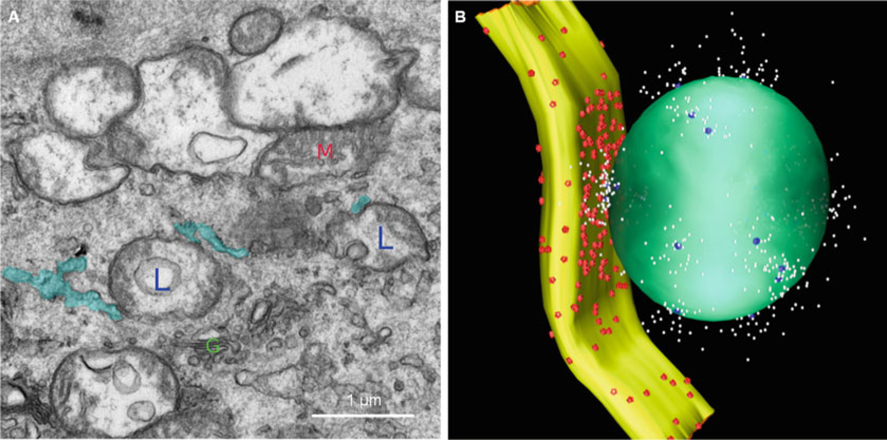
Fig. 13.2.

Lysosome–SR junction. The image obtained by electron microscopy shows the structure of the lysosome–SR junction. Lysosomes are labeled with L; SRs are labeled with blue color; the mitochondrion is labeled with M; the Golgi is labeled with G. Based on the dimensional characteristics of the lysosome–SR junction obtained by electron microscopy, we built a three-dimensional reconstruction of a typical lysosome–SR junction, including lysosome (green), SR (yellow), TRPML1 channel (blue), SERCA2 pumps (red), and Ca2+ (white)
