Fig. 5.
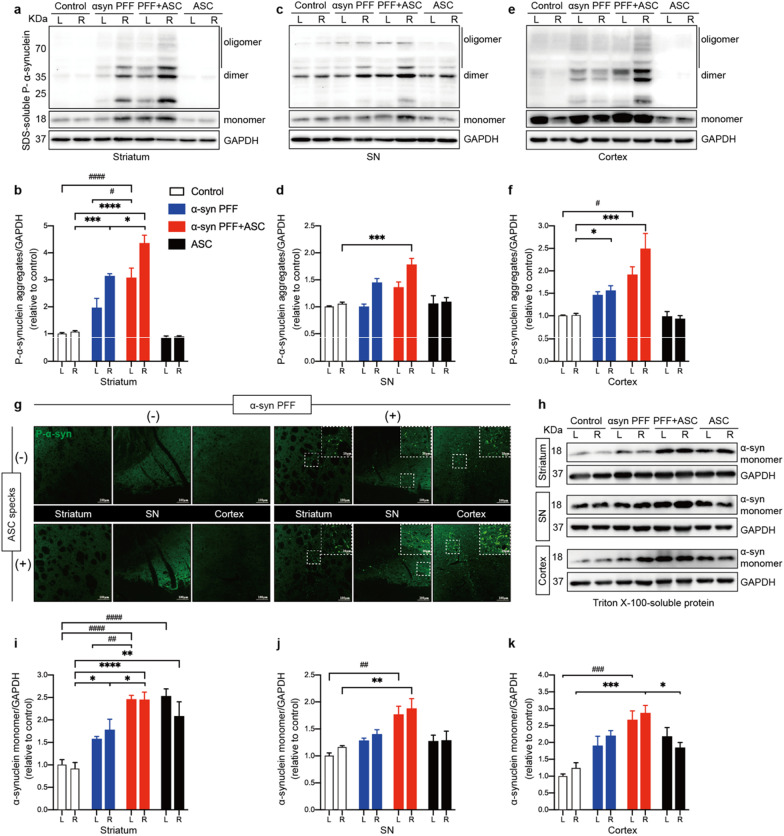
ASC specks exacerbated α-synuclein aggregation and propagation. a, b IB analysis and quantification of SDS-soluble phosphate-α-synuclein (P-α-syn) in bilateral striatum (n = 4 or 6). c, d IB analysis and quantification of SDS-soluble P-α-syn in bilateral SN tissues (n = 4 or 6). e, f IB analysis and quantification of SDS-soluble P-α-syn in cortex (n = 4 or 6). g Representative immunofluorescence staining of P-α-syn (green) in the ipsilateral striatum and SN regions of indicated mice. h–k IB analysis and quantification of triton-soluble α-synuclein monomers in the bilateral striatum, SN and cortex in indicated mice (n = 4 or 6). Data are presented as mean ± SEM and are analyzed by two-way ANOVA followed by Tukey’s post hoc test for multiple comparisons. Significance levels are: *p < 0.05, **p < 0.01, ***p < 0.001, ****p < 0.0001 (Right hemisphere of brain tissue); #p < 0.05; ##p < 0.01; ###p < 0.001; ####p < 0.0001 (Left hemisphere of brain tissue). Scale bars are as indicated. L left, R right, SN substantia nigra

