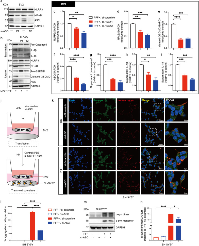
Fig. 7.
ASC knockdown suppressed PFFs-induced NLRP3 inflammasome activation in BV2 cells and upregulation of α-synuclein in SH-SY5Y cells. a IB analysis of NLRP3, NF-κB, ASC in mouse BV2 cells transfected with either si-scramble RNA or si-ASC RNA for 48 h. b–i IB analysis and quantification of NLRP3 inflammasome signals in BV2 cells challenged with 500 ng/ml LPS priming for 4 h followed by 1 μM PFFs stimulation for 16 h after transfection. j Schematic drawing of the BV2 cells and SH-SY5Y cells co-culture setup used in this study. Si-scramble RNA or si-ASC RNA transfected BV2 cells are treated with PBS or 1 μM PFFs and co-cultured with SH-SY5Y cells for 16 h. k Representative immunofluorescence images of P-α-syn (green) and human α-synuclein (red) in SH-SY5Y cells co-cultured with indicated-treated BV2 cells. The white dotted boxes in images are magnified on the right. l Quantification of the percentages of α-synuclein aggregates-positive cells per vision in SH-SY5Y cells. m, n IB analysis and quantification of α-synuclein in co-cultured SH-SY5Y cells. Data are obtained from at least three independent experiments. Data are presented as mean ± SEM and are analyzed by one-way ANOVA followed by Tukey’s post hoc test for multiple comparisons. Significance levels are: *p < 0.05, **p < 0.01, ***p < 0.001, ****p < 0.0001. Scale bars are as indicated

