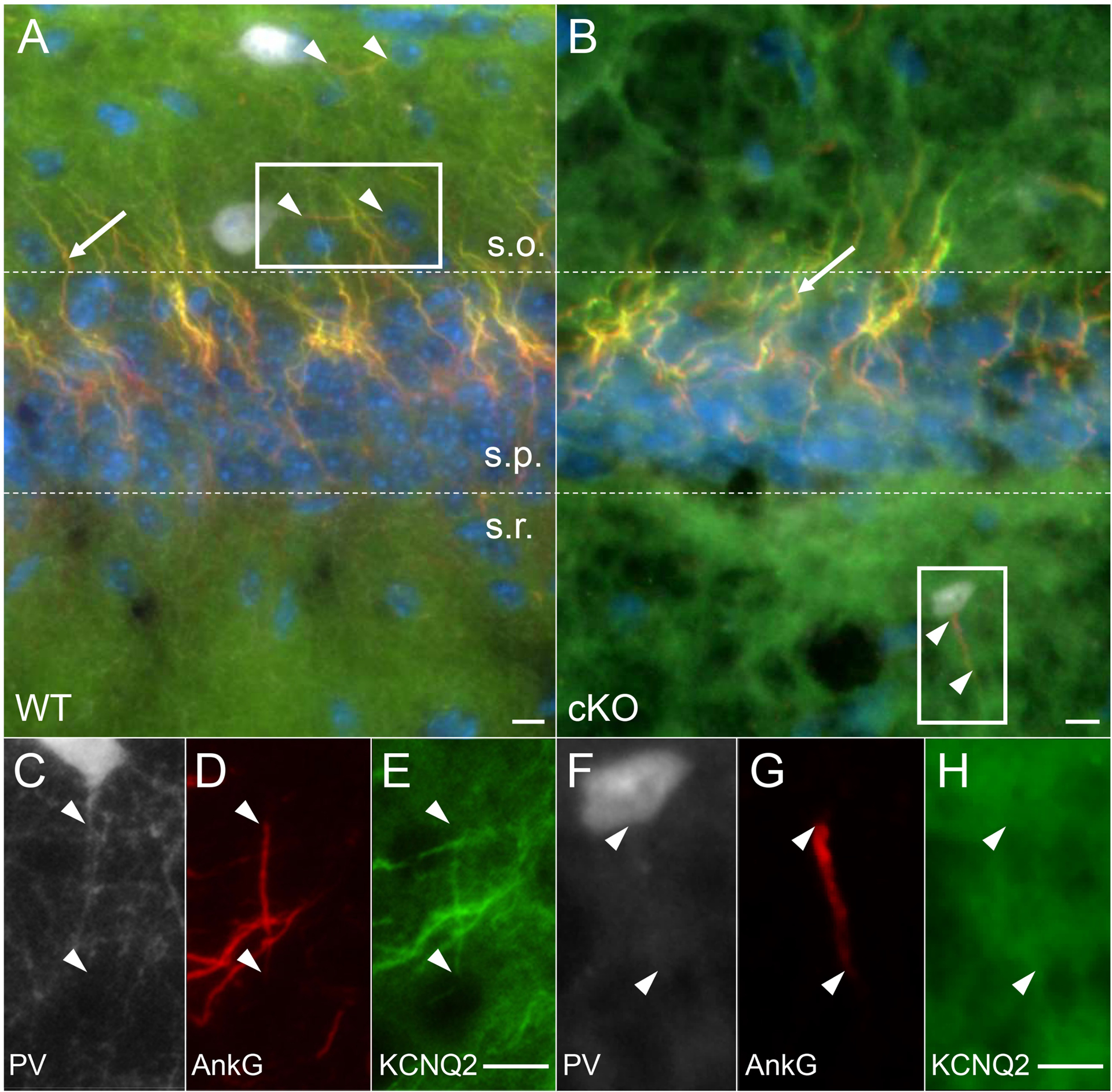
Figure 3. Selective deletion of KCNQ2 from CA 1 PV-IN in cKO mice.

(A) In WT mice, KCNQ2 (green) was co-expressed with AnkG (red, arrowhead) at the distal axon initial segment (AIS) of PV-IN (TdTomato – white) in stratum oriens (s.o.=stratum oriens, s.p.=stratum pyramidale, s.r.=stratum radiatum). (B) In cKO mice, KCNQ2 was no longer co-expressed with AnkG in PV-IN (here shown in stratum radiatum of CA1), but remained evident in the AIS of CA1 pyramidal cells in stratum pyramidale (arrow). (C-E) Inset from A, showing KCNQ2 expression in the distal 2/3 of the AIS. (F-H) Inset from B, showing absence of KCNQ2 immunoreactivity localized to the PV interneuron AIS (arrowheads showing beginning and end of AIS). Scales: 10 μm.
