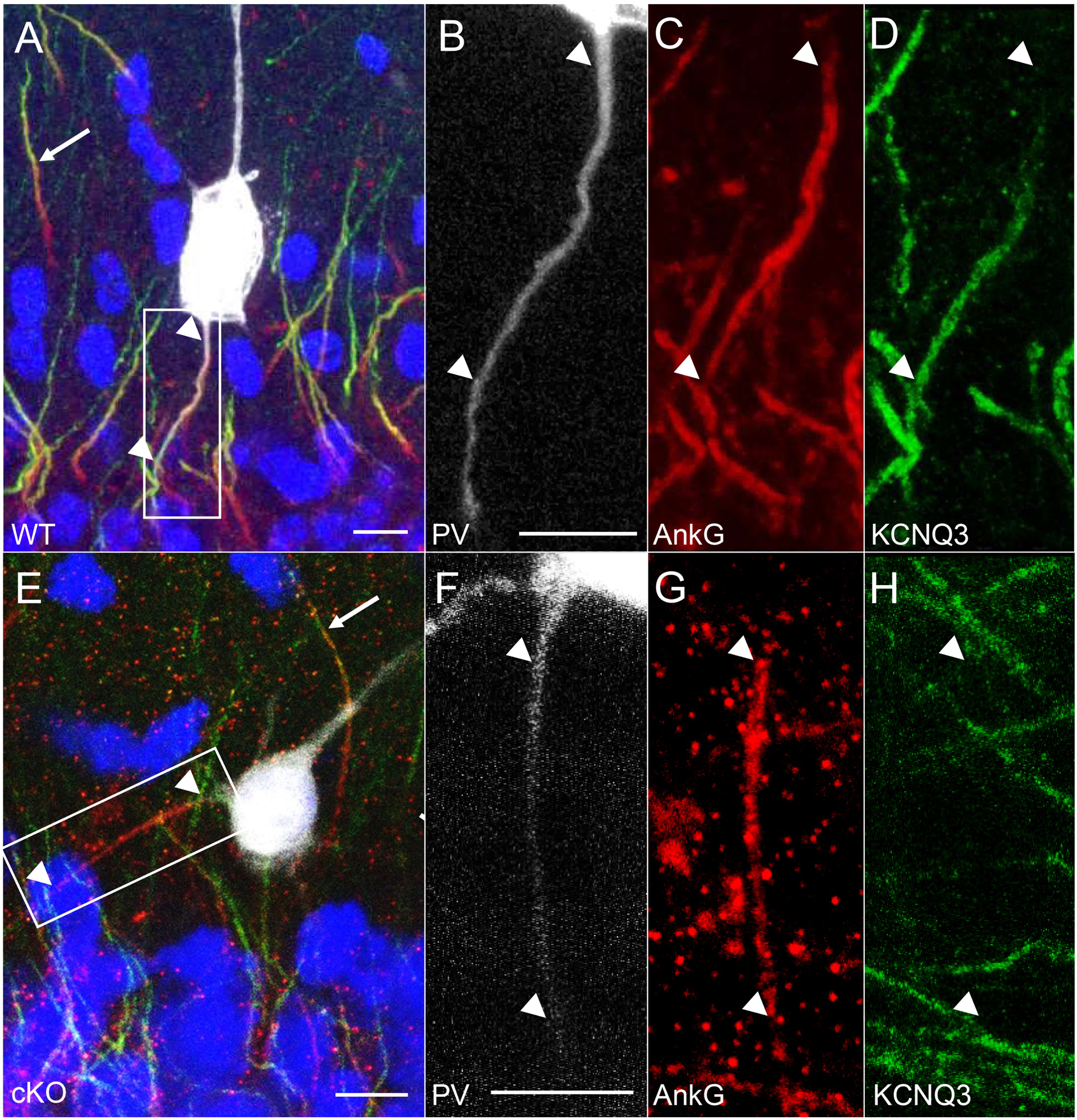
Figure 4. Absence of KCNQ3 AIS labeling in PV-INs in cKO mice.

(A-D) Representative images obtained with confocal imaging, where KCNQ3 (green) expression was seen at the distal AIS (arrowheads) labeled by AnkG (red) in a WT CA1 stratum pyramidale PV-IN, with non-PV-INs also showing distal AIS labeling of KCNQ3 in the opposite direction (arrow). (E-H) In a PV-IN from the cKO mouse, staining for KCNQ3 was absent in the AIS (arrowheads), but detected in the neighboring AIS of a non-PV-IN (arrow). Scale bars: 10 μm.
