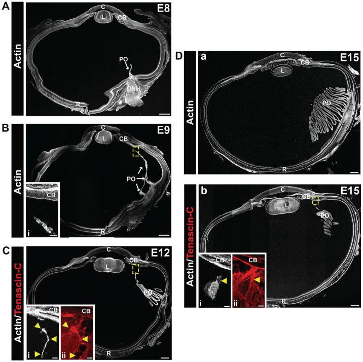
Figure 12.
Tenascin-C matrix fibrils link the developing vasculature of the pecten oculi with the ciliary body. Cryosections of E8, E9, E12, and E15 chick eyes were labeled with fluorescent-conjugated phalloidin (white) to visualize the development of the chick pecten oculi. At E8 (A), the pecten oculi is identified as a thin projection from the optic nerve head (ONH) into the vitreous. By E9 (B), the pecten oculi extends anteriorly through the vitreous to the ciliary body, the region in the dashed box shown at higher magnification as an inset (Bi). At E12 (C), the pleated structure of the pecten has begun to develop, and is more extensive by E15 (Da, b). Higher magnification insets in C and D are from consecutive sections that are also immmunolabeled for tenascin-C (Ci, Di—F-actin [white]; Cii, Dii—tenascin-C [red]). Tenascin C–rich fibrils are shown to provide a link between the pectin oculi and the ciliary body. Arrowheads in Ci and Di indicate the same positions in (Ci) or (Dbi). Mag. bars: (A) to (D) = 500 and 50 µm for the insets. C: cornea; CB: ciliary body; L: lens; PO: pecten oculi; R: retina. (A color version of this figure is available in the online journal.)

