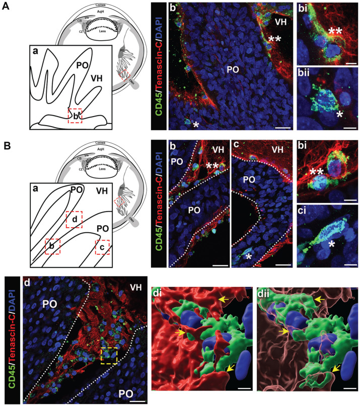
Figure 13.
Immune cells are highly concentrated in the vitreous matrix between the pleats of the pecten oculi. Cryosections of E12 chick eyes were co-immunolabeled with the pan-leukocyte marker CD45 (green). Nuclei were labeled with DAPI (blue). Confocal images were obtained in two regions as illustrated in the diagrams including the base of the optic nerve head (ONH) (Aa), and within the pleats (Ba) of the pecten oculi. Boxed in regions illustrate where each image was acquired. Immune cells are detected within the base of the ONH (Ab and Abii) and found entwined with tenascin-C fibrils on the outer edge of this structure (Ab and Abi). Immune cells were found to densely populate the pleated region of the pecten oculi, specifically the inner and outer surface of the folds (Bb, Bbi), in the matrix within the gaps between the folds (Bd), and more rarely within the fold itself (Bc, Bci). Migratory phenotype of immune cells entangled in matrix between folds is highlighted in 3D surface renderings of a confocal Z-stack from which (Bd) was taken with solid (Bdi) and transparent (Bdii) tenascin-C. Asterisks indicate immune cells at higher magnification from selected regions. Mag. Bars: Ab, Bb–d = 20 µm, Abi, Abii, Bbi, Bbii = 5 µm, Bdi, Bdii = 3 µm. PO, pecten oculi; ONH, optic nerve head; VH, vitreous humor. (Ab, Bb–c) Projection image; (Bd) single optical plane; (Bdi, Bdii) 3D surface structure. (A color version of this figure is available in the online journal.)

