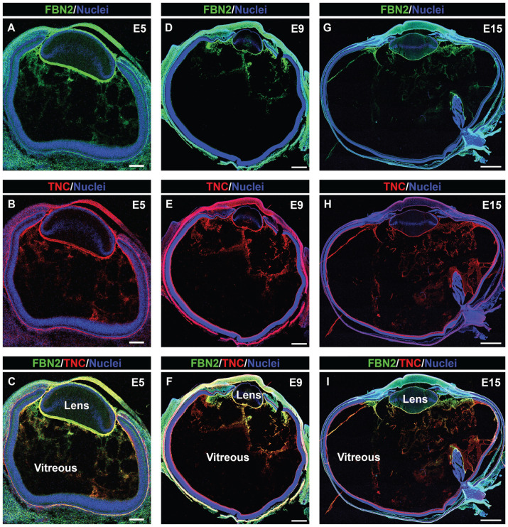
Figure 2.
Overview of tenascin-C/fibrillin-2 fibril organization during development of the chick embryo eye; 5× confocal tiles were acquired of cryosections of embryonic day 5 (A to C), 9 (D to F), and 15 (G to I) chick eyes that were co-immunolabeled for fibrillin-2 (green) and tenascin-C (red), and co-labeled for nuclei with DAPI (blue). Mag. bars: E5 = 100 µm, E9 = 500 µm, E15 = 1000 µm. (A color version of this figure is available in the online journal.)

