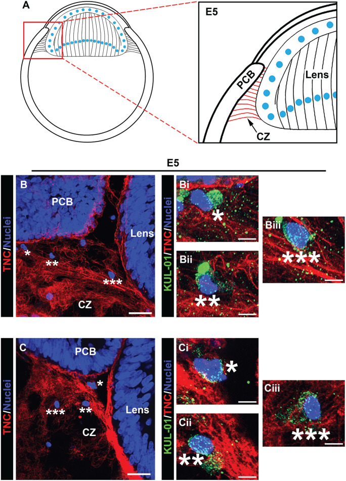
Figure 7.
Immune cells populate the ciliary zonules as early as E5. Cryosections of E5 heads were co-immunolabeled for tenascin-C (red) to highlight the ciliary zonules and KUL-01 (green), which is expressed by immune cells of the monocyte/macrophage lineage. Nuclei were labeled with DAPI (blue); 40× confocal z-stacks were acquired of the ciliary zonule region as identified in the diagram (A). Projection images at low magnification are shown with just the tenascin-C and DAPI labels (B, C) and the regions denoted by asterisks shown at high magnification including the KUL-01 labeling for the immune cells (Bi–iii; Ci–iii). Immune cells were identified along the zonule fibers. Mag. bars: (B) and (C) = 20 µm, (Bi–iii) and (Ci–iii) = 5 µm. (A color version of this figure is available in the online journal.)

