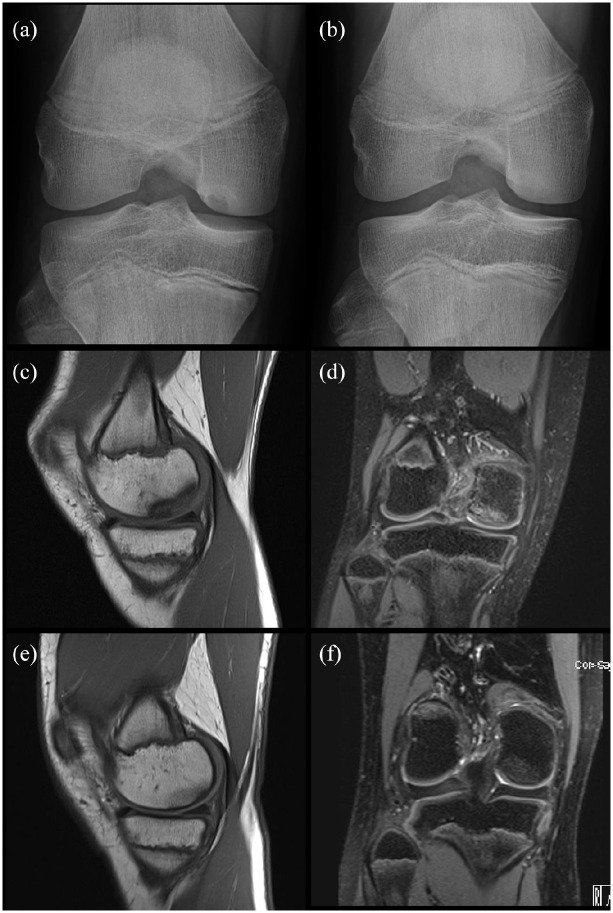
Figure 1.
Two cases of spontaneous healing of OCD. Case 1: 14 years old boy. X-ray notch view of the right knee (a) shows a stable OCD of the right medial femoral condyle. X-ray (b) after 6 months of sports restriction shows a complete spontaneous healing of OCD. Case 2: 12 years old, female. Detection of OCD on MRI after 18 months of knee pain. Coronal T1-weighted (c) and sagittal T2-weighted (d) MRI images show the area of OCD of the medial lateral condyle with low-signal intensity lesion on T1-images and a signal suggestive of bone marrow edema on T2-images. There is no sign of instability. Coronal T1-weighted (e) and sagittal T2-weighted (f) MRI images after 12 months of sporting activity restriction show OCD healing.

