
Figure 7. M1AP colocalizes with TEX11 and facilitates TEX11 recruitment at the recombination intermediates.
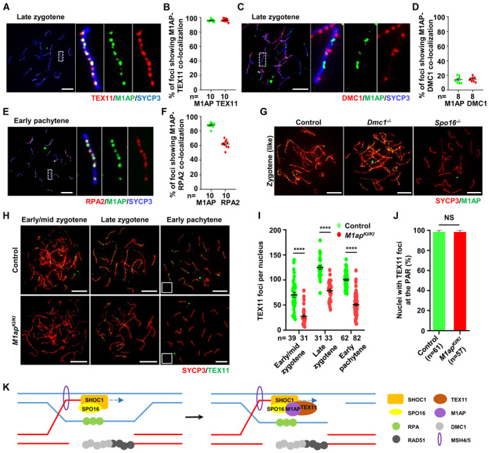
- Immunofluorescence staining of M1AP (green) and TEX11 (red) on the spermatocyte spreads of WT mice.
- Ratios of M1AP‐TEX11 co‐localizing foci to the M1AP total foci or TEX11 total foci at the indicated meiotic prophase substages. Each dot represents the ratio calculated from one nucleus.
- Immunofluorescence staining of M1AP (green) and DMC1 (red) on the spermatocyte spreads of WT mice.
- Ratios of M1AP‐DMC1 co‐localizing foci to the M1AP total foci or DMC1 total foci at the indicated meiotic prophase substages. Each dot represents the ratio calculated from one nucleus.
- Immunofluorescence staining of M1AP (green) and RPA2 (red) on the spermatocyte spreads of WT mice.
- Ratios of M1AP‐RPA2 co‐localizing foci to the M1AP total foci or RPA2 total foci at the indicated meiotic prophase substages. Each dot represents the ratio calculated from one nucleus.
- Immunofluorescence staining of M1AP (green) and SYCP3 (red) on spermatocyte spreads of control, Dmc1 −/− or Spo16 −/− mice.
- Immunofluorescence staining of TEX11 (green) and SYCP3 (red) on the spermatocyte spreads of control and M1ap KI/KI testes.
- The mean number of TEX11 foci per cell in control and M1ap KI/KI spermatocytes.
- Frequencies of nuclei with TEX11 foci detected at the PAR in spread early pachytene spermatocytes with touching XY chromosomes.
- Schematic diagram showing a proposed model in which M1AP acts as a partner of the ZZS complex in meiotic recombination in mouse meiotic prophase I. M1AP is recruited by SPO16 and facilitates TEX11 recruitment to stabilize recombination intermediates.
Data information: (A, C, E, G, and H) Scale bars, 10 μm. (B, D, F, I, and J) Data are presented as the mean ± SEM. n shows the number of cells scored from at least two biological replicates. (I and J) ****P < 0.0001; NS, not significant; two‐tailed Student's t‐test.