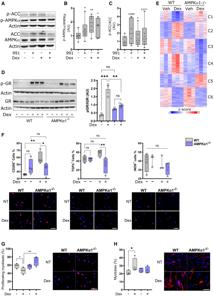
Figure 1. Glucocorticoid signaling through AMPK in macrophages.

-
ABone marrow‐derived macrophages (BMDMs) were treated with either the AMPK activator 991, Dexamethasone (Dex) or a combination for 1 h before analysis by Western blot.
-
B, CPhospho‐AMPK (B) and phospho‐ACC (C) were quantified.
-
DWT and AMPKα1−/− BMDMs were treated with Dex for 1 h before analysis by Western blot and GR phosphorylation at S211 was quantified.
-
EWT and AMPKα1−/− BMDMs were treated with Dex for 24 h and analyzed by RNA‐seq. Differentially expressed genes between any condition were clustered using c‐means clustering.
-
FWT and AMPKα1−/− BMDMs were treated with Dex for 24 h and anti−/pro‐inflammatory markers assessed using immunofluorescence.
-
G, HWT and AMPKα1−/− BMDMs were treated with Dex for 72 h, washed and conditioned medium was recovered after 24 h used to treat muscle stem cells. Proliferation (Ki67 immunostaining) (G) or differentiation (desmin immunostaining and counting the percentage of muscle cells with 2 nuclei or more) (H) were assessed in muscle stem cells.
Data information: results are means ± SEM or z‐score or displayed with boxes and whiskers in which the central band represents the median. N = 6 (B, C), 3 (D), 3–4 (F) of 4 biological replicates (G, H). *P < 0.05, **P < 0.01, ***P < 0.001 statistical analysis using Student's t‐test (B, C) or ANOVA tests (D, G, H). Bar = 25 μm (G, H).
Source data are available online for this figure.
