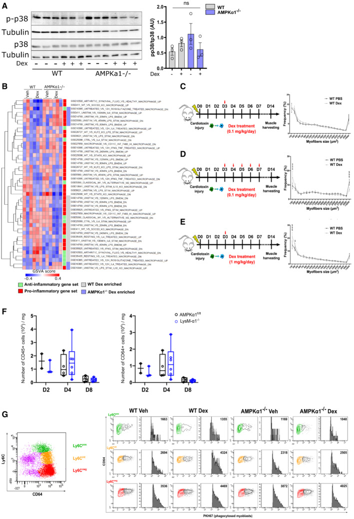
Figure EV1. Glucocorticoid treatment during skeletal muscle regeneration and signaling through AMPK in macrophages is independent of p38.

-
AWT and AMPKα1−/− Bone marrow‐derived macrophages (BMDMs) were treated with Dexamethasone (Dex) for 1 h before analysis by Western blot for p38 phosphorylation and quantified.
-
BRepresentation of GSVA analysis from Fig 1F with gene sets labeled.
-
C–EExperimental procedure of mice treatment with Dex. After cardiotoxin injection to damage the muscle, AMPKα1fl/fl (WT) and LysM‐α1−/− mice were treated with (C) a single dose of Dexamethasone (Dex) intra‐peritoneal (i.p.) (0.1 mg/kg) at day 3 (D3), or (D) multiple doses of Dex i.p. (0.1 mg/kg) on D3 to D7 after CTX, or (E) a single dose of Dex i.p. (1 mg/kg) at D3, and Tibialis Anterior (TA) muscles were harvested at day 8 (D8) and 14 (D14) after injury. Myofiber cross sectional area was then quantified.
-
FThe number of immune cells (CD45pos, left panel) and macrophages (CD45posCD64pos, right panel) per milligram of muscle tissue (TA muscle) was assessed by flow cytometry in AMPKα1fl/fl (WT) and LysM‐α1−/− mice at day 2, 4 and 8 after cardiotoxin injury.
-
GMuscles and cells were treated as described in the legend of Fig 2D. Plot on top shows the whole population of macrophages stained with CD64 and Ly6C. Plots on bottom show proportion of macrophages that have phagocytosed fluorescent dead myoblasts in the Ly6Cpos (green population), Ly6Cint (orange population) and Ly6Cneg (red population) macrophages in the various conditions.
Data information: results are means ± SEM (A) or box and whiskers in which the central band represents the median of three biological replicates (A) or 7 (D), 8–10 (C, E), 2–6 (F–G) animals. $ P < 0.05, $$$ P < 0.001 vs untreated WT, using ANOVA tests.
Source data are available online for this figure.
