
Figure 3. AMPKα1 in macrophages is required for glucorticoid‐dependent suppression of inflammation in endotoxin‐induced lung injury.
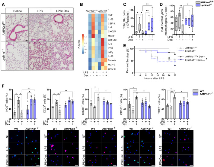
AMPKα1fl/fl (WT) and LysM‐α1−/− mice were treated with vehicle or with 10 mg/kg lipopolysaccharide (LPS) or LPS + Dexamethasone (Dex, 1 mg/kg), before an intravenous injection of oleic acid was made to induce lung inflammation.
-
AHematoxylin–eosin staining of lung tissue after 24 h.
-
BBroncho‐alveolar lavage (BAL) cytokine content was measured by Luminex after 24 h.
-
C, DAfter 24 h, mice were culled and cells infiltrating the lungs were analyzed. (C) The number of cells present in the BAL fluid was counted. (D) The number of pro‐inflammatory macrophages (F4/80pos Ly6Cpos) was evaluated by flow cytometry as a percentage of CD45pos cells.
-
ESurvival curve of mice treated with vehicle or Dex.
-
FMacrophage polarization was determined by immunofluorescence in vehicle, LPS and LPS + Dex treated WT and AMPKα1−/− BMDMs, data are expressed as percentage positive cells.
Data information: Results are displayed with box and whiskers in which the central band represents the median (C, D) or means ± SEM (F). N = 5–10 (C), 5–7 (D), 3–7 (data below detection threshold were excluded from analysis) (B), 13–15 (E) or 3 (F) biological replicates. Statistical analysis by ANOVA (C, D, F) or Gehan–Breslow–Wilcoxon test (E). *P < 0.05, **P < 0.01, ***P < 0.001. Bar = 20 μm (A), 25 μm (F).