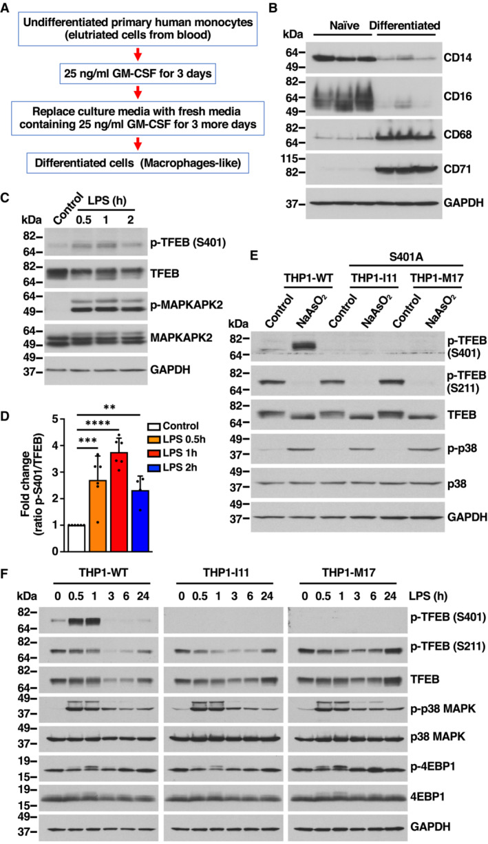
Figure EV3. TFEB‐S401 phosphorylation induced by NaAsO2 and LPS treatments is absent in S401A knock‐in THP1 cells. Related to Fig 3 .

- Flowchart indicating the different steps followed to differentiate primary human monocytes into macrophage cells.
- Immunoblot analysis of protein lysates from primary human monocytes undifferentiated (Naïve) or GM‐CSF‐differentiated primary macrophages. Samples are from three independent experiments.
- Immunoblot analysis of protein lysates from primary human macrophages incubated with 1 μg/ml LPS for the indicated times. Immunoblots are representative of at least four independent experiments.
- Quantification of immunoblot data shown in (C). Data are presented as mean ± SD using one‐way ANOVA (unpaired) followed by Dunnett's multiple comparisons test, **P < 0.01, ***P < 0.001 and ****P < 0.0001 as compared to untreated cells from at least four independent experiments.
- Immunoblot analysis of protein lysates from THP1 cells WT or TFEB‐S401A knock‐ins (clones I11 and M17) incubated with 100 μM NaAsO2 for 1 h.
- Immunoblot analysis of protein lysates from PMA‐differentiated THP1 cells WT or TFEB‐S401A knock‐ins (clones I11 and M17) incubated with 1 μg/ml LPS for the indicated times. Immunoblots are representative of at least three independent experiments.
Data information: n ≥ 3 biological replicates (each dot represents a biological replicate).
Source data are available online for this figure.
