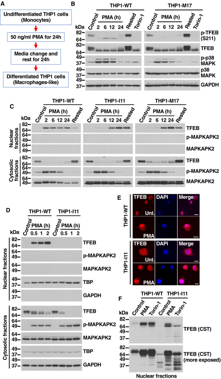
Figure 4. Activation of TFEB depends on S401 phosphorylation during macrophage differentiation.

- Flowchart indicating the different steps followed to differentiate Naïve into macrophage‐like THP1 cells.
- Immunoblot analysis of protein lysates from naïve THP1‐WT or TFEB‐S401A knock‐in (clone M17) cells treated with 50 ng/ml PMA for the indicated times, PMA‐differentiated THP1 (Rested) cells treated without or with 250 nM Torin‐1 for 1 h.
- Immunoblot analysis of proteins from nuclear and cytosolic fractions from naïve THP1‐WT or TFEB‐S401A knock‐in (clones I11 and M17) cells treated with 50 ng/ml PMA for the indicated times and PMA‐differentiated THP1 (Rested) cells.
- Immunoblot analysis of proteins from nuclear and cytosolic fractions from naïve THP1‐WT or TFEB‐S401A knock‐in (clone I11) cells treated with 50 ng/ml PMA for the indicated times.
- Immunofluorescence confocal microscopy of naïve THP1‐WT or TFEB‐S401A knock‐in (clone I11) cells treated with 50 ng/ml PMA for 1 h. Scale bars, 10 μm.
- Immunoblot analysis of proteins from nuclear fractions from naïve THP1‐WT or TFEB‐S401A knock‐in (clone I11) cells treated with 50 ng/ml PMA or 250 nM Torin‐1 for 1 h. Immunoblots are representative of at least three independent experiments. The antibody directed against the central region of TFEB was obtained from Cell Signaling Technology (CST).
Source data are available online for this figure.
