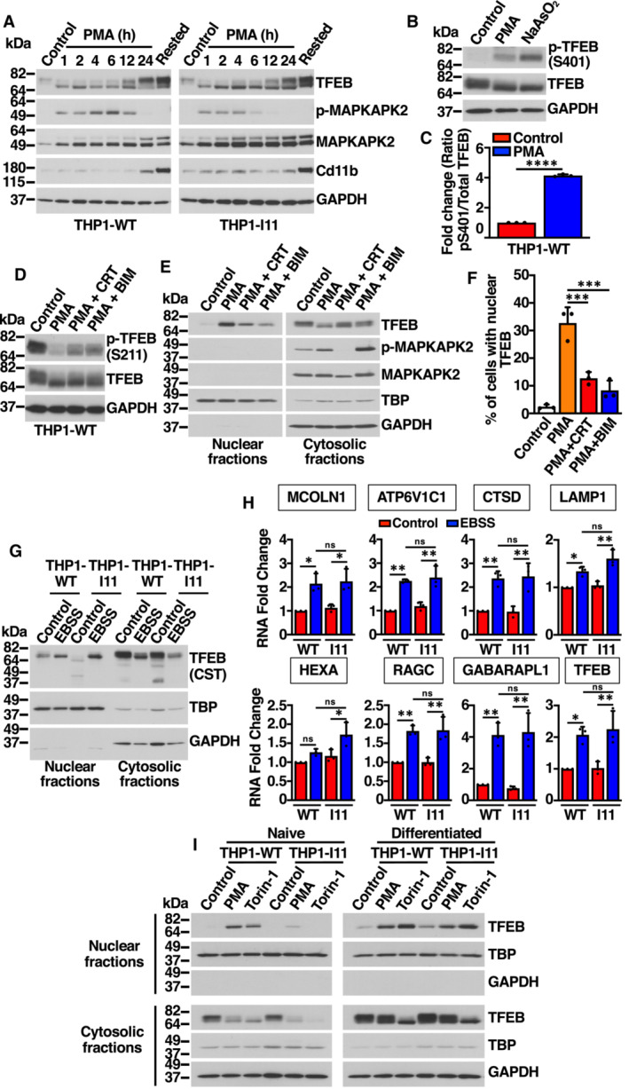
Figure EV4. PMA‐dependent activation of TFEB in THP1 cells. Related to Fig 4 .

- Immunoblot analysis of protein lysates from naïve THP1‐WT or TFEB‐S401A knock‐in (clone I11) cells treated with 50 ng/ml PMA for the indicated times, and PMA‐differentiated THP1 (Rested) cells.
- Immunoblot analysis of protein lysates from naïve THP1‐WT cells treated with 50 ng/ml PMA for 30 min and with 100 μM NaAsO2 for 1 h.
- Quantification of immunoblot data shown in (B). Data are presented as mean ± SD using unpaired Student's t‐test, ****P < 0.0001 from three independent experiments.
- Immunoblot analysis of protein lysates from naïve THP1‐WT cells treated with 5 μM CRT0066101(CRT, PKD inhibitor) or 5 μM Bisindolylmaleimide IV (BIS, PKC inhibitor) for 1 h prior to the addition of 50 ng/ml PMA for 1 h.
- Immunoblot analysis of proteins from nuclear and cytosolic fractions from naïve THP1‐WT treated with 5 μM CRT0066101(CRT, PKD inhibitor) or 5 μM Bisindolylmaleimide IV (BIS, PKC inhibitor) for 1 h prior to the addition of 50 ng/ml PMA for 1 h.
- Quantification of the nuclear localization of recombinant TFEB in HeLa cells expressing TFEB‐WT‐FLAG treated with 5 μM CRT0066101(CRT, PKD inhibitor) or 5 μM Bisindolylmaleimide IV (BIS, PKC inhibitor) for 1 h prior to the addition of 500 ng/ml PMA for 45 min. Data are presented as mean ± SD using one‐way ANOVA (unpaired) followed by Tukey's multiple comparisons test, ***P < 0.001 as compared to PMA‐treated cells, with > 200 cells counted per trial from three independent experiments.
- Immunoblot analysis of proteins from nuclear and cytosolic fractions from naïve THP1‐WT or TFEB‐S401A knock‐in (clone I11) cells nutrients starved with EBSS for 12 h. The antibody directed against the central region of TFEB was obtained from Cell Signaling Technology (CST).
- Relative quantitative RT–PCR analysis of the mRNA expression of lysosome‐ (MCOLN1, ATP6V1C1, CTSD, LAMP1, HEXA, RAGC) and autophagy‐related (GABARAPL1) genes in naïve THP1‐WT or TFEB‐S401A knock‐in (clone I11) cells nutrients starved with EBSS for 12 h. Data are presented as mean ± SD using one‐way ANOVA (unpaired) followed by Tukey's multiple comparisons test, *P < 0.05 and **P < 0.01 from three independent experiments.
- Immunoblot analysis of proteins from nuclear and cytosolic fractions from naïve THP1‐WT or TFEB‐S401A knock‐in (clone I11) cells or PMA‐differentiated THP1 (Rested) cells treated with either 50 ng/ml PMA or 250 nM Torin‐1 for 1 h.
Data information: n = 3 biological replicates (each dot represents a biological replicate).
Source data are available online for this figure.
