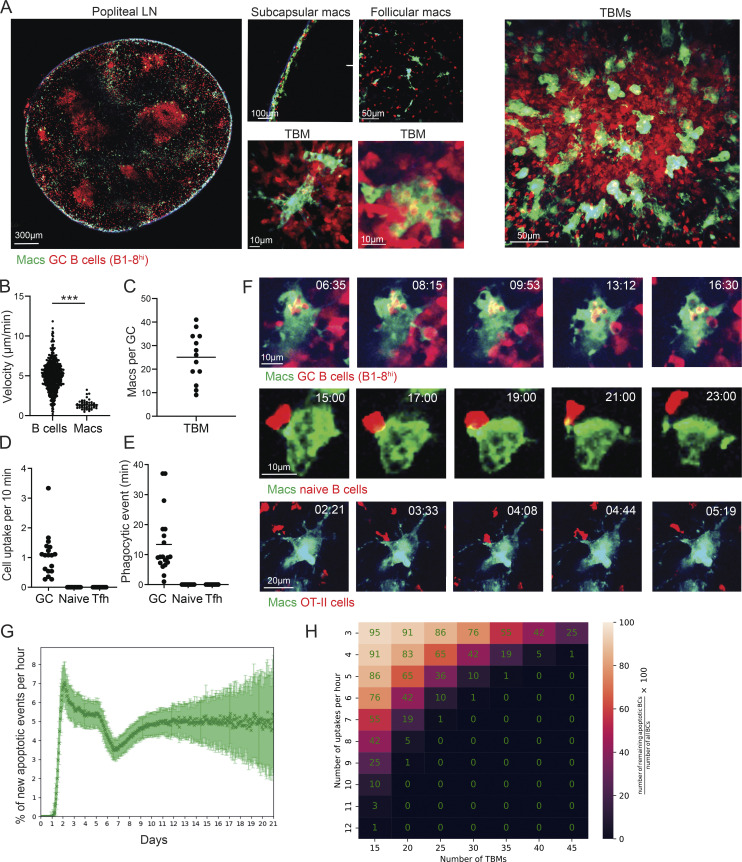
Figure 1.
B cell disposal in GCs is mediated by TBMs through highly dynamic dendrites. (A) TPLSM images of popliteal LNs of OVA-primed CX3CR1GFP mice that received TdTomato+ B1-8hi B cells, 7 d after intra-footpad NP-OVA boost. (B) Velocity of B cells and TBMs in the GC. Each dot represents a single cell. (C) Number of macrophages in the GCs. Each dot represents a GC; three mice were imaged. (D and E) OVA-primed CX3CR1GFP mice that received either TdTomato+ B1-8hi B cells prior to boosting, naive B cells 1 d before TPLSM intravital imaging, or TdTomato+ OT-II T cells prior to initial immunization. TPLSM intravital imaging was performed 7 d after intra-footpad boost with NP-OVA. Number of cells (D) and cell uptake rate per single TBM (E). Each dot represents a single TBM uptake event. The data were obtained from two to three mice. Two-tailed Student’s t test; ***, P < 0.0001; ns, not significant. (F) TPLSM images of popliteal LNs of CX3CR1GFP interactions with the transferred lymphocytes. Two to three mice were imaged per condition. (G) Simulation of apoptotic B cell number per hour based on previously published GC dynamic modeling. (H) Prediction of the number of macrophages required for removal of all of the apoptotic cells in the GC. Number of apoptotic cells = (number of TBMs × uptake rate)/the number of B cells in the GC.

