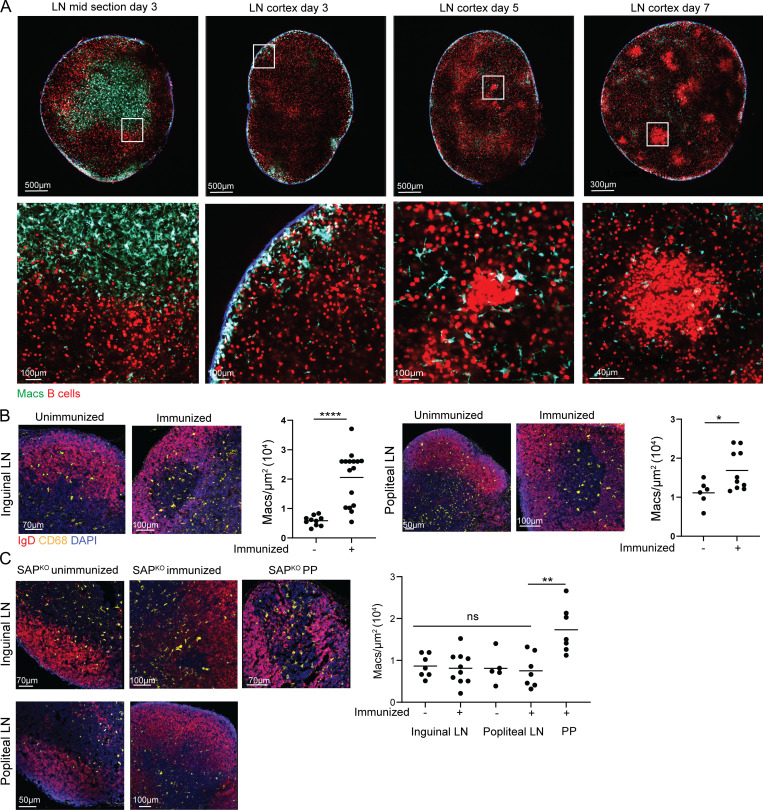
Figure 2.
Macrophage infiltration into LN follicles depends on GCs. (A) TPLSM images of popliteal LNs derived from CX3CR1GFP mice that were adoptively transferred with TdTomato+ B1-8hi B cells and imaged 3, 5, and 7 d after intra-footpad immunization with NP-OVA. Each time point was repeated twice, with two mice in each repeat. (B) Immunofluorescence staining of LN-derived from immunized and unimmunized mice. Each dot represents the density of macrophages in LN follicles; three to four mice were analyzed from each group. (C) Immunofluorescence staining of LNs derived from unimmunized and immunized SAPKO mice. Each dot represents the density of macrophages in the follicles of three mice. Line represents the mean. *, P < 0.05; *, P < 0.01; ****, P < 0.0001; ns, not significant. Two-tailed Student’s t test in B, one-way ANOVA in C.

