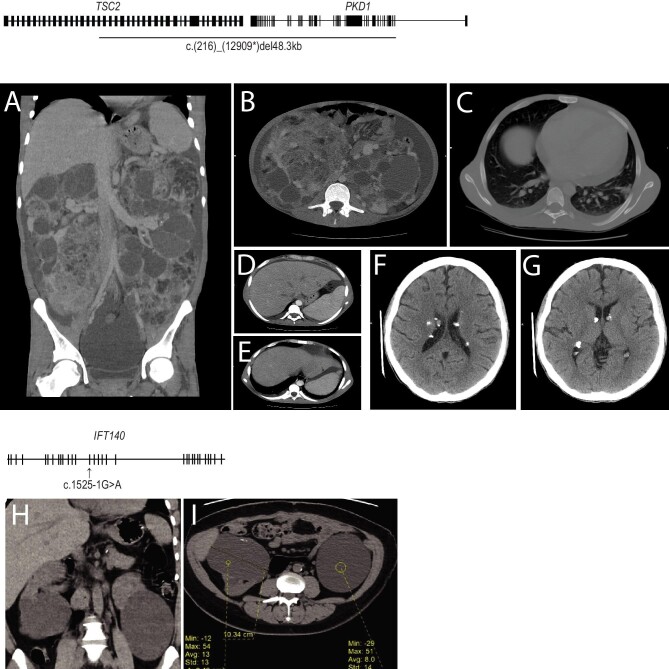
Figure 3:
Diagnostic imaging of two rare PKD cases. (A) A CT scan of a patient with TSC2/PKD1 contiguous gene syndrome shows markedly enlarged kidneys with innumerable variable-sized cortical and parenchymal cysts, some showing scattered tiny mural calcifications. (B) Renal masses of mixed soft tissue and fat attenuation, suggestive of angiomyolipoma, are noted with a large amount of ascites. (C) A CT scan shows bilateral basal lung bronchiectatic changes along with atelectatic bands. (D and E) Small left hepatic lobe cysts were noted. (F and G) Multiple calcified subependymal nodules were noted. (H and I) A CT scan of the IFT140 patient shows enlarged kidneys by the presence of multiple variable-sized cortical and to lesser extent parenchymal renal cysts. (H) The largest cysts are the lower polar exophyting ones. (I) Small mural calcifications are seen at the right-sided lower polar cyst.

