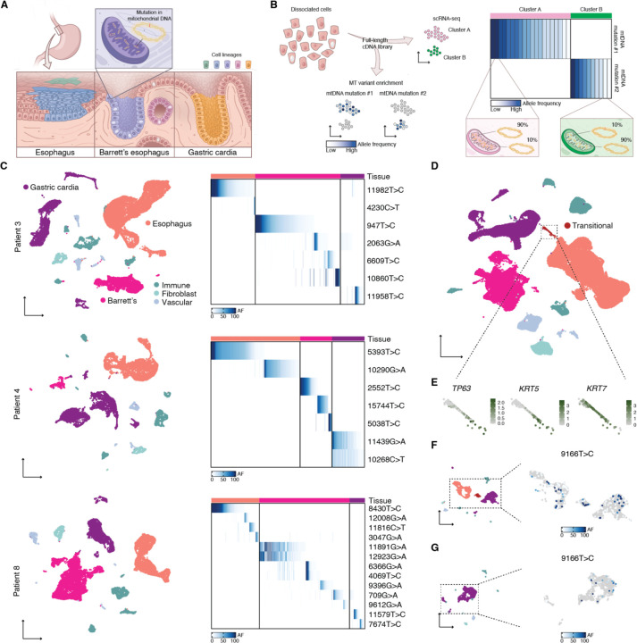
Fig. 1. Cell lineages in human gastroesophageal junction tissues labeled by mtDNA mutations.
(A) Barrett’s esophagus occurs at the gastroesophageal junction between esophageal squamous and gastric cardia tissues. Clones within these tissues can be traced using distinctive mtDNA mutations. (B) Conventional scRNA-seq libraries can be enriched for mtDNA mutations, enabling the linking of clones to cell states. (C) UMAPs of scRNA-seq of matching Barrett’s esophagus, esophageal squamous, and gastric cardia tissues for three Barrett’s esophagus patients; tissues contained supporting immune, fibroblast, and vascular cells. Adjacent heatmaps show the allele frequencies (AF) of mtDNA mutations within Barrett’s esophagus, esophageal, and gastric cardia cells. (D) UMAP of scRNA-seq of all the samples collected in this study; highlighted are transitional basal progenitor cells from the normal squamocolumnar junction. (E) Callouts of the transitional cells from (D) featuring the expression of basal progenitor markers. (F) UMAP of scRNA-seq of a single squamocolumnar junction biopsy from patient 9; in the callout is the same UMAP, colored with the allele frequency of mutation 9166T>C. (G) UMAP of scRNA-seq of a single gastric cardia biopsy from patient 9; in the callout is the same UMAP, colored with the allele frequency of mutation 9166T>C.

