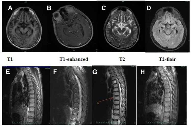
Figure 2.
(A–D) Brain MRI on day 5 of onset revealed multiple enhanced lesions on the bilateral perimesencephalic cistern, sylvian fissure and cerebral longitudinal fissure and its peripheral cerebral cortex, as well as the frontal lobe and parietal lobe; (E–H) MRI of the thoracic segment on day 14 of onset revealed the 2–4, 12 thoracic medullary enlargements, with longer T1, T2 signals, and spinal cord membrane thickening with cystic image, which were enhanced.

