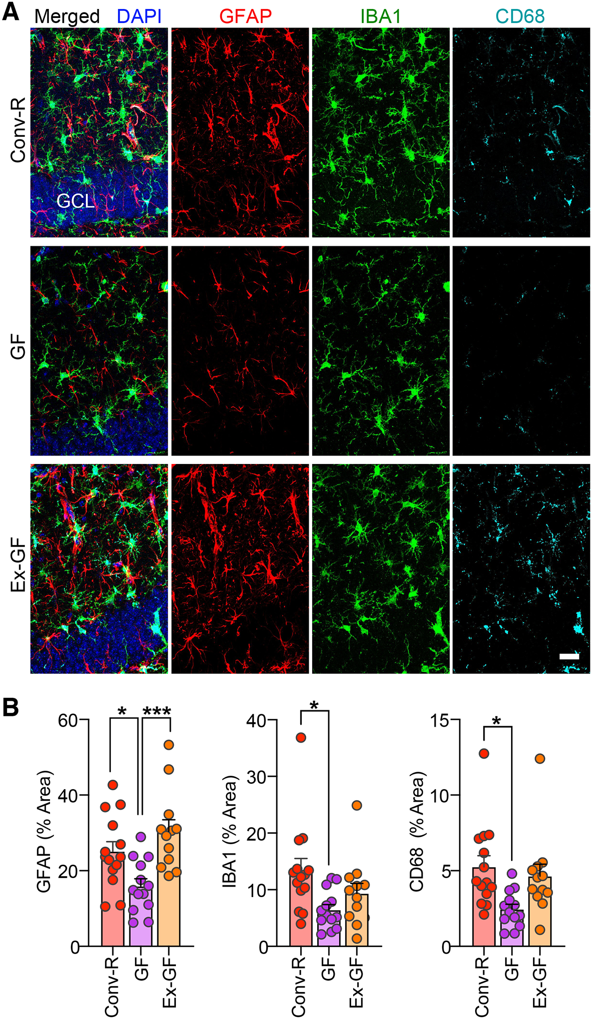
Fig. 2. Germ-free TE4 mice exhibit reduced reactive gliosis.

(A) Representative immunofluorescence images of hippocampal sections from 40-week-old male Conv-R, GF, and Ex-GF mice stained with antibodies to GFAP (red), Iba-1 (green), and CD68 (cyan), as well as DAPI (blue). Scale bar, 25 μm. GCL, granule cell layer. (B) Percent of the area of sections taken from the hippocampus covered by GFAP (left), Iba-1 (middle), CD68 (right) staining. Mean values ± SEM are shown. (n=12–14/group). Statistical significance was defined by one-way ANOVA with Tukey’s post-hoc test. *, p < 0.05, ***, p < 0.001. (See Table S1 for full statistical results).
