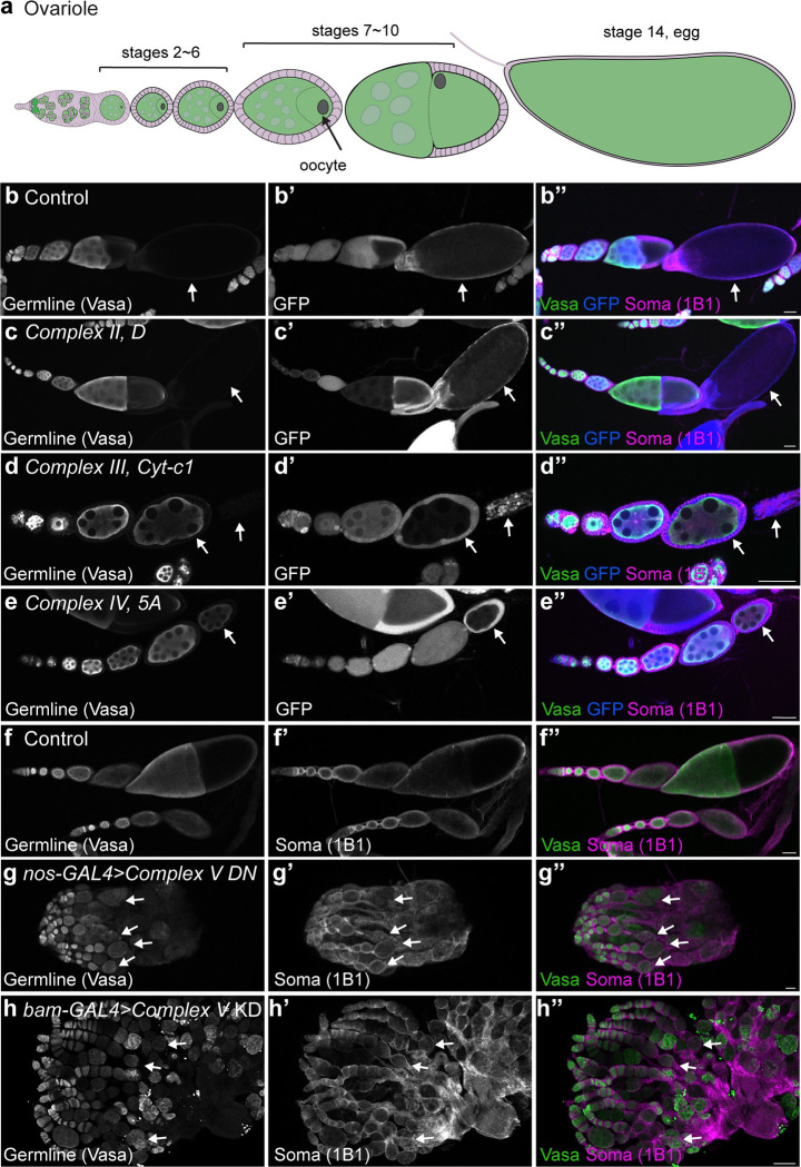
Fig 2. Egg development requires mitochondrial oxidative phosphorylation.
(a) Schematic of an ovariole. An egg chamber is comprised of one oocyte, fifteen nurse cells and surrounding somatic follicle cells. Oogenesis comprises 14 stages resulting in the formation of a mature egg. (b-e) Representative images of Control (b), Complex II subunit D (c), Complex III Cyt-c1 (d) and Complex IV subunit 5A (e) mosaic ovarioles 14-days post-clone induction. Arrows indicate GFP-negative mutant cells. (f, g) Representative images of 2–3 day old Control (f) and germline expressed (nos-GAL4) Complex V dominant negative (DN) (g) in heterozygous CVc deficiency ovaries. (h) Representative image of a 2–3 day old bam-GAL4 driven CVα RNAi ovary. Arrows indicate the last stage observed in the representative ovariole. For all images scale bars represent 100 μm. For exact genotypes see S2 Table.

